Quantcast
Channel: airway Archives - emDOCs.net - Emergency Medicine Education
Viewing all 46 articles
Browse latest View live

Intubating the Critically Ill Patient

$
0
0

Intro / Main Questions

The following is a review of the most recent literature regarding intubation of critically ill patients. Specifically, the following questions will be addressed:

What is the safest way to intubate a hypotensive patient?

Which patients are at risk for peri-intubation hypotension and cardiac arrest?

What other special circumstances should be considered in intubating critically ill patients?

Recap Basics

  • Prep: Assess for difficult airway, have backup plan
  • Pre-oxygenation: NRB vs NIPPV or manual ventilation (if SpO2 <91%) and passive O2 w/ 5L NC.
  • Pre-treatment (optional): Atropine for <1yr, lidocaine for reactive airway and increased ICP, fentanyl for increased ICP and CV emergency
  • Induction: Etomidate, ketamine, propofol, thiopental, or midazolam/fentanyl
  • Paralysis: Succinylcholine 45 sec, rocuronuim 60 sec
  • Placement: Most skilled personnel, minimize # of attempts
  • Post procedure: Lung-protective vent settings, confirm placement, ABG, HOB to 30-45 to improve lung mechanics, in-line suction, NG/OG, humidify air, sedation/analgesia

What’s New

Cardiac arrest complicating endotracheal intubation (ETI)1

Two independent variables associated with post-ETI arrest:

  1. Pre-induction shock index (SI = HR/SBP) > 0.9: OR increases 1.16 for every 0.1 increase in SI
  2. Weight: OR increases 1.37 for every 10kg increase in weight

Predicting post-intubation hypotension (PIH)2

  • SI > 0.8 associated with increased risk
  • PIH associated with increased in-hospital mortality

How to intubate the hypotensive patient3

  • Pre-treat scopolamine if able: 0.4mg IVP induces amnesia, decreases secretions
  • Have crystalloid (sepsis) or blood/FFP (trauma) infusing
  • Norepi BEFORE intubation to get MAP >80, can use PIV until CVC can be placed post-intubation
  • Have bolus dose pressor ready: epi 5mcg 1:100,000 (not phenylephrine)
  • Midazolam/fentanyl takes too long to act (3-5 min); don’t use it for induction
  • Decrease dose of induction agent: e.g. 10% of normal propofol dose or middle ground dosing ketamine 0.25-0.5 mg/kg, but actually need more of etomidate
  • Paralytic is cardiac output-dependent, therefore pt in shock requires higher dose: succinylcholine 2mg/kg or rocuronium 1.6mg/kg
  • Vent: low and slow (+pressure will drop BP); low PS/PEEP, start tidal volume (Vt) 6ml/kg

Delayed Sequence Intubation (DSI)3

  • For delirious patient with hypoxia, this is alternative to bagging the patient
  • Requires having vent immediately available
  • Ketamine (1-2mg/kg then aliquots of 0.5mg/kg) to disassociate patient
  • Ketamine keeps airway reflexes and does not suppress respirations
  • Vomiting happens during EMERGENCE, not sedation
  • Wait 3 minutes while patient breathes through NRB or NIPPV (if not saturating well on NRB then has shunt physiology and needs PEEP)
  • Paralytic then wait 45 seconds
  • Intubate

Avoiding complications during ETI/PPV4

Hypotension
  • Local anesthesia only if possible or
  • Small titrated dose of sedative (0.3mg/kg propofol based on ideal weight)
  • Begin volume resuscitation before ETI
  • Have pressor ready
  • Start w/ PEEP 5 (0 for COPD) and Vt = 6 ml/kg then titrate up for plateau pressure (Pplt) = 20-30
Acid-base complications
  • Avoid hypotension as above
  • Choose initial setting similar to the pt’s respiratory status prior to ETI (but RR <30 to avoid auto-PEEP)
  • ABG in 15 minutes
  • If complicated by seizure (lactic acidosis) hyperventilate 25-30/min and give bicarb if unstable or insufficient time to document acidosis as cause
Asthmatic
  • Permissive hypercapnea
  • Reduce RR
  • Reduce Vt
  • Reduce iTime / increased expiratory time (promote full exhalation)
  • Keep Pplt <30cm H2O
  • May need sedation / muscle relaxation to decrease RR
Dyspnea / hypoxemia despite high FiO2 (ARDS)
  • Begin w/ Vt 6ml/kg
  • Titrate Vt to maintain Pplt <30
  • Incr PEEP by 3-5 q2-3min and decr Vt to keep Pplt 25-30 until SpO2 >90% on FiO2 = 60%
  • Ensure patient-ventilator synchrony
Traumatic Brain Injury
  • Pre-treatment optional (not evidence-based): Lidocaine 1.5 mg/kg 3 min prior then fentanyl 3 mcg/kg if not hypotensive
  • Induction: Etomidate 0.3 mg/kg or ketamine 1-2 mg/kg if hypo or normotensive
  • Paralysis: Succinylcholine preferred (rocuronium is ok too)
  • Hyperventilate temporarily if patient continues to deteriorate after osmotic agents

Bottom Line / Pearls & Pitfalls

  • BVM with PEEP valve and NC at 10L is “poor man” CPAP
  • Don’t bag before or in between attempts if SpO2 >90%
  • All induction agents can induce hypotension, but ketamine generally causes a sympathetic “surge” with middle ground dosing
  • Be wary of ketamine in patients with CAD, cardiac emergency, hypertension and tachycardia
  • Absolute contraindications to ketamine: age <3months and history of schizophrenia
  • Shock patient requires lower dose of induction agent (except etomidate) and higher dose of paralytic
  • Succinylcholine contraindications: FH of malignant hyperthermia, hyperkalemia, burns > 24h, crush injuries > 3 days, sepsis after 7 days, congenital / acquired myopathies, denervation illness, chronic neuropathy
  • Apply pressure to cricoid, not the thyroid cartilage for Sellick
  • Start low and slow with the vent, +pressure will drop BP even further
  • Have post-intubation checklist

Sources / Further Reading

  1. Heffner A, et al. Incidence and factors associated with cardiac arrest complicating emergency airway management. Resuscitation 84 (2013) 1500-1504.
  2. Heffner A, et al. Predictors of the complication of postintubation hypotension during emergency airway management. Journal of Critical Care (2012) 27, 587–593.
  3. Weingart S. Preoxygenation, Reoxygenation, and Delayed Sequence Intubation in the Emergency Department. The Journal of Emergency Medicine April 2010.
  4. Manthous, CA. Avoiding Circulatory Complications During Endotracheal Intubation and Initiation of Positive Pressure Ventilation. The Journal of Emergency Medicine, Vol. 38, No. 5, pp. 622–631, 2010.
  5. Griesdale D, et al. Airway Management in Critically Ill Patients. Lung (2011) 189:181–192.
  6. Heffner A, et al. The frequency and significance of postintubation hypotension during emergency airway management.  Journal of Critical Care (2012) 27, 417.e9-417.e13.
  7. Reynolds S. Airway Management of the Critically Ill Patient Rapid-Sequence Intubation. Chest Vol 127, Issue 4 (April 2005).
  8. Green SM, et al. Clinical Practice Guideline for Emergency Department Ketamine Dissociative Sedation: 2011 Update. Ann Emerg Med. 2011;57:449-461.
  9. Sehdev RS, et al. Ketamine for rapid sequence intubation in patients with head injury in the emergency department. Emergency Medicine Australasia (2006) 18, 37–44.
  10. Price B, et al. Hemodynamic consequences of ketamine vs etomidate for endotracheal intubation in the air medical setting. American Journal of Emergency Medicine 31 (2013) 1124–1132.
  11. Robinson N. In patients with head injury undergoing rapid sequence intubation, does pretreatment with intravenous lignocaine/lidocaine lead to an improved neurological outcome? A review of the literature. Emerg Med J 2001;18:453–457.
  12. Orebaugh SL. Succinylcholine: Adverse Effects and Alternatives in Emergency Medicine. Am J Emerg Med 1999;17:715-721.
  13. MacLennan N. Anesthesia for Major Thermal Injury. Anesthesiology 1998; 89:749-70.

Discussion Questions / Future Exploration

  1. Therapeutic intervention aimed at optimizing pre-intubation hemodynamics
  2. Optimal drugs for intubating hemodynamically unstable patients
  3. Optimal drug combinations for intubation of other subsets of critically ill patients
Edited by Alex Koyfman

The post Intubating the Critically Ill Patient appeared first on emdocs.


Supraglottic Airway Review

$
0
0

Background

As of 2008, the Nation Emergency Medical Services Information System (NEMSIS) data from 16 states reported 4 million EMS activations with over 88,000 airway interventions ranging from bag valve mask to cricothyroidotomy.1  Although the bag valve mask and endotracheal tube remain the cornerstones of airway management, many alternatives have emerged in prehospital care.  Availability of airway adjuncts will depend upon training and local department purchasing.  This review will discuss the basics of extraglottic airway adjuncts available for prehospital airway management in adults and pediatrics.

Extraglottic devices, or supraglottic airways as they are commonly called, insert through the patient’s oropharynx and temporarily manage ventilation.  The devices do not pass through the vocal cords and only act as a temporary, not definitive, airway.  Some devices have both supraglottic and infraglottic structures, making the name extraglottic the most encompassing term.  Aspiration of gastric contents can occur while the devices are in place, making the endotracheal tube (ETT) the only definitive airway.

The Laryngeal Mask Airway (LMA)

In 1981, Dr. Archie Brain invented the first extraglottic airway device, the LMA Classic.  The cLMA, was first sold in the United Kingdom in 1988 and then in the United States in 1992 by LMA North America.  The cLMA, a reusable device, exists for prehospital care as the LMA-Unique (disposable), LMA Fastrach (disposable with ability to intubate through), LMA ProSeal (additional gastric suction port), and LMA Supreme (Proseal plus biteblock).  When inserted the LMA follows the path of a food bolus moving along the hard palate to the hypopharynx and then to the esophagus.  Finally the LMA sits with the distal tip posterior to cricoid cartilage and masks the glottis for ventilation.2

lma
Figure 1: Placement of the LMA from a dorsal view of pharynx (Image Courtesy of LMA North America Inc.)


A meta-analysis of randomized trials in 1994 compared the LMA to the ETT and BVM.  Compared to BVM, a LMA was easier to place by new trainees, and improved oxygen saturations.  Compared to an ETT, the LMA increased speed of placement but the ETT demonstrated higher seal pressure and less gastric insufflation.3

In pediatrics the LMA also serves as a well documented rescue device for “can’t intubate, can’t ventilate” situations.  Sizes for the LMA range from 1kg neonates to >70kg patients.4

Esophageal Tracheal Combitube (ETC)

Described in 1987, by Dr. Frass in Austria, the ETC, or commonly called the Combitube, functioned as a resuscitation rescue device from the beginning.  Unlike the LMA which anesthesiologist used before prehospital providers, the Combitube focused primary on the prehospital and emergency environment.  The Combitube, is a disposable double-lumen double cuffed device commonly inserted into the patient’s esophagus but 5% of the time inserted into the trachea.  The provider blindly inserts the device and either via auscultation or capnometry confirms placement in the esophagus or trachea by ventilating trough one of the two ports.  For tracheal intubation ventilation occurs via the more distal lumen and for esophageal placement ventilation occurs via the proximal lumen.

The major criticism of this device stems from the multi-lumen design.  In stressful emergent situations prehospital providers must spend time determining the correct port for ventilation.5 The Combitube has no place in pediatric airway management since Covidien only produces two sizes for adults.

Laryngeal Tube (LT)

VBM Medizintechnik GmbH in Germany in 1999, created the LT in response to criticism regarding the multi-lumen difficulties of the Combitube.  The LT gained in popularity in the United States for prehospital use when   King Systems produced a disposable version in 2003.  The LT by design eliminated the 5% of tracheal intubations that occurred when using the Combitube freeing prehospital providers from deciding which port to use for ventilation.  The LT has an oropharyngeal end esophageal inflatable cuff and a single lumen ventilation lumen.  Both cuffs inflate with one port.6  The LT constantly achieves esophageal intubation and but if tracheal intubation were to occur the provider could not ventilate the patient.7 Novice providers have also demonstrated rapid insertion times for the LT half as long as tradition ETT placement.8  King Systems currently produces a size two LT which accommodates patients as small as 12kg.  Smaller pediatric disposable LT sizes have not been manufactured in the United States.

LMA Fastrach

fastrach
Figure 4: LMA Fastrach (Image Courtesy of LMA North America Inc.)

Stemming from the success of the LMA classic, Dr. Brain developed the Fastrach LMA (FT-LMA) to allow for blind intubation through the device.  The FT-LMA, also referred to as the intubating LMA, has a larger internal diameter to accommodate a proprietary ETT, a rigid airway tube, an epiglottic elevating bar, and a tracheal tube guiding ramp.9 A multicenter study demonstrated  a blind intubation success rate with the special manufacturer ETT of 96.2%.10 The FT-LMA does not accommodate sizes for pediatric use.

I-Gel

i-gel
In 2003, in the UK, Intersurgical Ltd. developed the i-gel.  The device similar to the LMA differs mainly by the lack of an inflatable laryngeal mask.  Intersurgical manufactures the device from thermoplastic elastomer to conform precisely to the laryngeal and laryngeal anatomy.  The device in theory should provide a greater seal pressure and increased speed of insertion by not requiring inflation.12  A recent study on hands off time in CPR during a cardiac arrest did not demonstrate a time savings using the i-gel vs. other extraglottic devices.13  The i-gel similar to the LMA supports sizes down to neonates.

Air-Q

In 2004, Dr. Daniel Cook, founder of Cookgas developed an alternative to the FT-LMA.  The Air-Q, similar to the FT-LMA allows for blind intubation once the device has been placed.  Unlike the FT-LMA the air-Q has a shorter and larger bore ventilating lumen with a removable ventilation connector.  The modifications were designed to eliminate the difficulty many providers experienced when intubating through the FT-LMA.  A shorter ventilation lumen should allow easier removal of the device over the ETT once in place.  Also the air-Q does not require a proprietary ETT, but supports the standard polyvinyl chloride tube found in most intubation kits.11  The air-Q supports sizes down to neonates.

Current Controversies

Within the last 10 years large scale prehospital trials have focused on neurologic outcomes in out of hospital cardiac arrest (OHCA) among different airway interventions.  It is not enough to have return of spontaneous circulation if there is significant morbidity as a result.  In Japan, a nationwide observational study of all out-of-hospital cardiac arrest between 2005-2007 demonstrated slightly poorer one-month neurologic outcomes in the patients whose airways were managed with extraglottic devices.  Neurologically favorable one month survival was associated with slightly, but significantly, poorer neurological outcomes compared with tracheal intubation.15  A more recent study with a greater number of OHCAs in Japan from 2005-2010 demonstrated that any ETT, ETC, LMA, or LT was associated with a decreased neurologically favorable survival.16  For pediatrics (age ≤12) no clear neurologic or survival benefit has been demonstrated between ETI and BVM in prehospital airway management.13

Overview

With a multitude of extraglottic devices on the market the LMA and LT remain the most common in the prehospital setting due to simplicity of use and reliability.  For pediatric use, the LMA has the largest range of sizes and clinical literature backing its use, although no large prehospital studies have been performed.  BVM should still function as the best and most reliable means of ventilating a child in the prehospital environment.19 Future extraglottic devices for prehospital use may include the FT-LMA and Air-Q.  Both allow the option to attempt blind intubation through the device.  No studies have demonstrated benefits of converting from an extraglottic device to an ETT.  None of the new devices such as FT-LMA, i-gel, or air-Q, have any adult or pediatric prehospital data to support their widespread adoption. For austere environments the Combitube requires the most force to dislodge (28.3lbs of force) while the King is most easily dislodged (12.5lbs of force).  The Army equips field medics with LTs and Combitubes.  Just as with endotracheal intubation proficiency and successful placement requires continue practice on patients and during simulation.  Lastly, 2010 American Heart Association recommend chest compressions take priority over establishing a patients airway as to minimize hands off time during cardiac arrest.

References / Further Reading

  1. Wang HE, Mann NC, Mears G, Jacobson K, Yealy DM. Out-of-hospital airway management in the United States. Resuscitation. 2011:378–385. http://www.ncbi.nlm.nih.gov/pubmed/21288624
  2. Laryngeal Mask Airway. Published Dec. 2012. Accessed  Dec. 2012 http://emedicine.medscape.com/article/82527-overview
  3. Brimacombe JR. The advantages of the LMA over the tracheal tube or facemask: a meta-analysis. Can J Anaesth. 1995;42(11):1017–1023. http://www.ncbi.nlm.nih.gov/pubmed/8590490
  4. Pediatric Anesthesia. Accessed Dec. 2012. http://openanesthesia.org/index.php?title=Pediatric_Anesthesia
  5. Combitube – Airway Device Difficult Airway Tutorial. Created Feb. 2007. Accessed Dec. 2012. http://vam.anest.ufl.edu/airwaydevice/combitube/index.html
  6. Emergency Airway Management: Other Supraglottic devices. Accessed Dec 2012.
  7. http://www.airwaycam.com/other-devices.html
  8. Russi CS, Wilcox CL, House HR. The laryngeal tube device: a simple and timely adjunct to airway management. Am J Emerg Med. 2007;25(3):263–267. http://ems.pgpic.com/pdf/kingairway_airway_management_independent.pdf
  9. Brain AI, Verghese C, Addy EV, Kapila A. The intubating laryngeal mask. I: Development of a new device for intubation of the trachea. British Journal of Anaesthesia. 1997;79(6):699–703. http://bja.oxfordjournals.org/content/79/6/699.full.pdf
  10. Baskett PJ, Parr MJ, Nolan JP. The intubating laryngeal mask. Results of a multicentre trial with experience of 500 cases. Anaesthesia. 1998;53(12):1174–1179. http://onlinelibrary.wiley.com/doi/10.1046/j.1365-2044.1998.00614.x/abstract
  11. Emergency Airway Management: Laryngeal Masks Accessed Dec. 2012 http://www.airwaycam.com/Supraglottic-airways.html
  12. Ruetzler K, Gruber C, Nabecker S, et al. Hands-off time during insertion of six airway devices during cardiopulmonary resuscitation: a randomised manikin trial. Resuscitation. 2011;82(8):1060–1063. http://www.resuscitationjournal.com/article/S0300-9572(11)00221-8/abstract
  13. Gausche M, Lewis RJ, Stratton SJ, et al. Effect of out-of-hospital pediatric endotracheal intubation on survival and neurological outcome: a controlled clinical trial. JAMA. 2000;283(6):783–790. http://jama.jamanetwork.com/article.aspx?articleid=192366
  14. Highlights of the 2010 American Heart Association Guidelines for CPR and ECC. Accessed Dec. 2012. http://www.heart.org/idc/groups/heart-public/@wcm/@ecc/documents/downloadable/ucm_317350.pdf
  15. Tanabe S, Ogawa T, Akahane M, et al. Comparison of Neurological Outcome between Tracheal Intubation and Supraglottic Airway Device Insertion of Out-of-hospital Cardiac Arrest Patients: A Nationwide, Population-based, Observational Study. J Emerg Med. 2012:1–9.
  16. Hasegawa K, Hiraide A, Chang Y, Brown DFM. Association of prehospital advanced airway management with neurologic outcome and survival in patients with out-of-hospital cardiac arrest. JAMA. 2013;309(3):257–266. doi:10.1001/jama.2012.187612.
Edited by Manpreet Singh

The post Supraglottic Airway Review appeared first on emdocs.

Airway Subtleties in Critically Ill Patients

$
0
0

Airway Subtleties in Critically Ill Patients: Salicylate Poisoning, Hypotensive/Septic, and Obtunded DKA

image1
‘Fish in the oropharynx.’ Sim training on Kangaroo Island, rural Australia via KIDocs.org. Reposted with permission (thanks!).

Salicylate-Poisoned Patients

When salicylate-poisoned patients are ventilated using standard settings you may harm your patients by diminishing the respiratory alkalosis, which facilitates the passage of salicylate into the CNS. Also the use of sedatives and paralytics can result in CO2 retention and respiratory acidosis, which can further facilitate the shift of salicylate into the CNS.

Because of this many toxicologists believe that intubation should be avoided if possible and performed only if the patient has true respiratory failure (worsening acidosis, hypoxemia). Endotracheal intubation and mechanical ventilation can be associated with rapid worsening of clinical salicylate toxicity and increased mortality unless a normal or slightly alkalemic blood pH is maintained via hyperventilation and achievement of a low PCO2 and/or intravenous sodium bicarbonate.

The following are recommendations for salicylate-poisoned patients based on a retrospective case series using the New York City Poison Control Center (NYCPCC) database for cases of salicylate poisoning, defined as a peak serum concentration >50 mg/dL, who had mechanical ventilation:

  • Avoid intubation if possible. Intubation should only be performed if patient truly has respiratory failure (worsening acidosis, hypoxemia).
  • Ensure that alkalinization of plasma and urine are initiated early and prior to intubation if possible.
  • Avoid paralytics and high doses of sedatives during rapid sequence intubation. Try to minimize the time that patient’s ventilatory drive is compromised.
  • Place an arterial line for frequent blood gas monitoring.
  • Frequent blood gas monitoring to ensure that an appropriately high minute ventilation is achieved. The goal is to maintain an arterial pH of 7.5–7.6.
  • Consider pressure-controlled ventilation. Adjust the rate to obtain the desired minute ventilation. This will allow delivery of maximal tidal volumes while controlling peak airway pressures. Any mode can be used as long as physiologic goals are being met. Adjust the settings based on the arterial blood gas to achieve goal pH.
  • Monitor closely for ‘‘breath-stacking’’ and ventilator asynchrony due to tachypnea.
  • Collaboration with intensivist recommended.

Further Reading

  1. American College of Medical Toxicology, Guidance Document: Management Priorities in Salicylate Toxicity, Available at: http://www.acmt.net/cgi/page.cgi/zine_service.html?aid=4210&zine=show
  2. Stolbach AI, Hoffman RS, Nelson LS. Mechanical ventilation was associated with acidemia in a case series of salicylate-poisoned patients. Acad Emerg Med 2008;15(9):866-9.
  3. http://www.ncbi.nlm.nih.gov/pubmed/12705252

Sepsis-Induced ARDS

Sepsis is the leading cause of death in U.S. hospitals, affecting 750,000 Americans and killing between 28 and 50 percent of those people each year. Managing the airway and ventilation settings in a patient with sepsis-induced ARDS can be challenging. The following are guidelines from the Surviving Sepsis Campaign, a joint collaboration of the Society of Critical Care Medicine and the European Society of Intensive Care Medicine committed to reducing mortality from severe sepsis and septic shock worldwide.

Mechanical Ventilation

  • Target a tidal volume of 6 mL/kg predicted body weight in patients with sepsis-induced ARDS (grade 1A vs. 12 mL/kg).
  • Plateau pressures should be measured in patients with ARDS and initial upper limit goal in a passively inflated lung be ≤ 30 cm H2O (grade 1B).
  • Positive end-expiratory pressure (PEEP) should be applied to avoid alveolar collapse at end expiration (atelectotrauma) (grade 1B).
  • Strategies based on higher rather than lower levels of PEEP should be used for patients with sepsis-induced moderate or severe ARDS (grade 2C).
  • Recruitment maneuvers should be used in sepsis patients with severe refractory hypoxemia (grade 2C).
  • Prone positioning should be used in sepsis-induced ARDS patients with a PaO2/FiO2 ratio ≤100 mm Hg in facilities that have experience with such practices (grade 2B).
  • Mechanically ventilated sepsis patients should be maintained with the head of the bed elevated to 30-45 degrees to limit aspiration risk and to prevent the development of ventilator-associated pneumonia (grade 1B).
  • Noninvasive mask ventilation (NIV) should be used in that minority of sepsis-induced ARDS patients in whom the benefits of NIV have been carefully considered and are thought to outweigh the risks (grade 2B).
  • A weaning protocol should be in place and that mechanically ventilated patients with severe sepsis undergo spontaneous breathing trials regularly to evaluate the ability to discontinue mechanical ventilation when they satisfy the following criteria: a) arousable; b) hemodynamically stable (without vasopressor agents); c) no new potentially serious conditions; d) low ventilator and end-expiratory pressure requirements; and e) low FiO2 requirements which can be met safely delivered with a face mask or nasal cannula. If the spontaneous breathing trial is successful, consideration should be given for extubation (grade 1A).
  • Against the routine use of the pulmonary artery catheter for patients with sepsis-induced ARDS (grade 1A).
  • A conservative rather than liberal fluid strategy for patients with established sepsis-induced ARDS who do not have evidence of tissue hypoperfusion (grade 1C).

Sedation, Analgesia, and Neuromuscular Blockade in Sepsis

  • Continuous or intermittent sedation should be minimized in mechanically ventilated sepsis patients, targeting specific titration endpoints (grade 1B).
  • Neuromuscular blocking agents (NMBAs) should be avoided if possible in the septic patient without ARDS due to the risk of prolonged neuromuscular blockade following discontinuation. If NMBAs must be maintained, either intermittent bolus as required or continuous infusion with train-of-four monitoring of the depth of blockade should be used (grade 1C).
  • A short course of NMBA of not greater than 48 hours for patients with early sepsis-induced ARDS and a PaO2/FiO2 < 150 mm Hg (grade 2C).

Use of Etomidate

The Surviving Sepsis Campaign Guidelines also discourage the use of etomidate if adrenal suppression is suspected. “Although the clinical significance is not clear, it is now recognized that etomidate, when used for induction for intubation, will suppress the hypothalamic-pituitary-adrenal axis. Moreover, a subanalysis of the CORTICUS trial revealed that the use of etomidate before application of low-dose steroids was associated with an increased 28-day mortality rate. An inappropriately low random cortisol level (< 18μg/dL) in a patient with shock would be considered an indication for steroid therapy along traditional adrenal insufficiency guidelines.”

Further Reading

  1. Surviving Sepsis Campaign, International Guidelines for Management of Severe Sepsis and Septic Shock. Crit Care Med. 2013 Feb;41(2):580-637. Available at http://www.sccm.org/Documents/SSC-Guidelines.pdf
  2. http://www.ncbi.nlm.nih.gov/pubmed/23903852
  3. http://www.ncbi.nlm.nih.gov/pubmed/23859579
  4. http://www.ncbi.nlm.nih.gov/pubmed/21394520

Diabetic Ketoacidosis with Severe Metabolic Acidosis

Diabetic Ketoacidosis is very common and is responsible for more than 500,000 hospital days per year at an estimated cost of 2.4 billion USD. Patients in DKA can look terrible. They may be obtunded, hypotensive, have respiratory fatigue after compensating for a while with Kussmaul breaths, and may have underlying sepsis. Their electrolytes are usually off and they can have severe metabolic acidosis. Patients are usually breathing at a maximum for respiratory compensation and trying to intubate them using standard approaches can cause a period of apnea which can kill your patient who is dependent on that respiratory compensation.

Fluid Therapy

The ADA recommends a gradual correction of glucose and osmolality as well as the judicious use of isotonic or hypotonic saline, depending on serum sodium and the hemodynamic status of the patient. In the absence of cardiac compromise, isotonic saline (0.9% NaCl) is infused at a rate of 15–20 ml/kg during the first hour. Subsequent choice for fluid replacement depends on hemodynamics, the state of hydration, serum electrolyte levels, and urine output. In general, 0.45% NaCl infused at 250–500 ml/h is appropriate if the corrected serum sodium is normal or elevated; 0.9% NaCl at a similar rate is appropriate if corrected serum sodium is low.  Once the plasma glucose is 200 mg/dl, 5% dextrose should be added to replacement fluids to allow continued insulin administration until ketonemia is controlled while at the same time avoiding hypoglycemia.

VBG vs ABG

You can feel confident sending off a VBG instead of an ABG. A VBG gives you the same information as an ABG except for the PO2 and O2 sat, but we have our monitors that we can use instead to give us this information (EMRAP May 2013).

Electrolyte Repletion

After IVF the next step in management is electrolyte repletion, especially potassium. Repleting electrolytes should be done prior to starting a patient on an insulin drip.  The ADA recommends that potassium replacement should begin with fluid therapy, and insulin treatment should be delayed until potassium concentration is restored to >3.3 mEq/l to avoid life-threatening arrhythmias and respiratory muscle weakness. The ADA also recommends repleting phosphate to avoid cardiac and skeletal muscle weakness and respiratory depression due to hypophosphatemia.

Sodium Bicarbonate

The use of sodium bicarbonate is controversial. Studies have shown that it may have little effect on patient outcome and can actually cause harm by causing increased risk of hypokalemia, decreased tissue oxygen uptake, cerebral edema, and development of paradoxical central nervous system acidosis. The ADA recommends that adult patients with a pH <6.9 should receive 100 mmol sodium bicarbonate (two ampules) in 400 ml sterile water (an isotonic solution) with 20 mEq KCI administered at a rate of 200 ml/h for 2 h until the venous pH is >7.0. If the pH is still <7.0 after this is infused, they recommend repeating infusion every 2 h until pH reaches >7.0. Some believe that giving sodium bicarbonate won’t work as patients are already breathing at their maximum. Unless they blow off the bicarb-generated CO2, they won’t increase their pH significantly (EMCrit Episode 3; EMRAP May 2013).

Insulin Bolus vs Insulin Drip

The ADA recommends an insulin bolus for adults, but no bolus in pediatrics when you start an insulin infusion. Some recommend not giving the bolus since studies have shown that patients are more likely to have hypoglycemic episodes when started with a bolus and have no difference in the change of anion gap (EMRAP May 2013).

RSI Medications

In cases requiring intubation, the ADA recommends that the paralytic succinylcholine should not be used if hyperkalemia is suspected; it may acutely further elevate potassium.

Airway Pearls

In Scott Weingart’s EMCrit Episode 3 he describes his method for intubating a DKA patient with severe metabolic acidosis. For preoxygenation he recommends using the “pseudo-NIV technique,” which is placing a NIV mask on the patient and connecting it to a ventilator on SIMV mode to allow the patient to take spontaneous breaths. Below are notes from his podcast.

Vent Settings during Preoxygenation

  • Mode Volume SIMV
  • Vt 550 ml
  • FiO2 100%
  • Flow Rate 30 lpm “aim nice, slow breaths, over 1 second (on normal ventilator setting usually less <1sec)”
  • PSV 5-10
  • PEEP 5
  • RR 0 (most important part, pt breaths on their own)

Getting ready to intubate

  • Attach ETCO2 and observe value (try and keep this value at all times)
  • Push the RSI Meds
  • Turn the Resp Rate to 12 (note we are giving breaths at all times so there is no apneic period)
  • Perform jaw thrust (while the pt is still wearing the BiPAP mask)
  • Wait 45 seconds. This violates the tenets of RSI, but keeping the pt alive is probably more crucial right now.
  • Most experienced operator should intubate the patient (first pass success most important).
  • Attach the ventilator
  • Confirm tube placement by observing ETCO2
  • Immediately increase Respiratory Rate to 30
  • Change Vt to 8 cc/kg predicted IBW
  • Change Flow Rate to 60 lpm, this is the normal setting for intubated patients

Why 30 BPM? To maintain eucapnea (normal capnea) you need 60 cc/kg/min. After intubation you need 120 cc/kg/min (because of additional dead space) to stay at a PCO2 of 40. But we want a PCO2 of at least 20 so we need to double it so 240 cc/kg/min, which when divided by 8 cc/kg comes out to 30 BPM to get the tidal volume we need.

  • Make sure ETCO2 is at least as low as it was when you started
  • Check blood gas (check pH and pCO2)
  • Pat yourself on the back

Further Reading

  1. www.EMCrit.org, Episode 3, Intubating the Patient With Severe Metabolic Acidosis. Available at http://emcrit.org/podcasts/tube-severe-acidosis/
  2. www.EMRAP.org, May 2013 Episode, DKA Myths
  3. American Diabetic Association Consensus Statement, Kitabchi A, et al. Hyperglycemic Crises in Adult Patients With Diabetes. Diabetes Care. 2009 July; 32(7): 1335–1343. Available at http://www.ncbi.nlm.nih.gov/pmc/articles/PMC2699725/
  4. Viallon A et al. Does bicarbonate therapy improve the management of severe diabetic ketoacidosis? Crit Care Med. 1999 Dec;27(12):2690-3.
  5. http://www.ncbi.nlm.nih.gov/pubmed/?term=%22DKA%22+%22intubation%22+%22emergency+medicine%22
  6. http://www.ncbi.nlm.nih.gov/pubmed/16911005
Edited by Alex Koyfman

The post Airway Subtleties in Critically Ill Patients appeared first on emdocs.

Yellowstone Advanced Airway Course Pearls

$
0
0

Thanks to Twitter I was privileged to attend a free advanced airway course hosted by Dr. Richard Levitan a few weeks ago.  This is an incredible 3-day course set in the beautiful Tetons near Yellowstone National Park. The course features lectures, Q&As, and hands-on stations to improve your airway assessment and intubation skills.

Instead of summarizing the entire course, because you really have to experience it yourself to get the full effect, I’m writing down the top 10 pearls I took away from this phenomenal experience.

  1. Intubate one step at a time. Instead of ramming the blade in the airway, take a deep breath and approach the airway in a step-wise fashion. First, find the uvula. Second, perform epiglottoscopy! Track down the uvula until you find the epiglottis. Third, remove the epiglottis off the posterior wall (suction may help remove that sticky film keeping the epiglottis on the posterior wall). Then laryngoscopy, to find a landmark (cords, notch, arytenoids). Finally, place the tube.
  2. Take time to position your patients. Everyone, regardless of age or size, should have ear-to-sternal notch. Want to avoid laundry? Try this.
    yellowstone1
  3. Learn the airway anatomy. Don’t rely on 2D pictures to understand the airway. Look at the CT imaging of your patients and learn how the turbinates are positioned and where the epiglottis rests in comparison to the hyoid. Understanding positioning and location will improve your skills.
  4. The floor of the nose is flat, when inserting anything into the nose, always stay flat and go straight back.yellowstone2
  5. Levitan has amazing recommendations for performing a cricothyrotomy. He emphasizes making a vertical incision BEFORE going horizontal. This procedure is a bloody mess and very tactile, you want to feel your landmarks before making each cut.
  6. When it comes to a cricothyrotomy, don’t rely on fine motor skills to find the landmarks. You will be too nervous and your hands won’t stay still. Avoid using only one fingertip to palpate and use your entire hand with the laryngeal handshake.
  7. Sedate your patients before invasive procedures to attenuate recall and PTSD.
  8. Learn how to manage yourself, a team, and a patient in a crisis and avoid fixation error. Biggest lesson was his focus on what to do after an unsuccessful intubation attempt; do not try the same method twice. Use bougie, use new blade size, or different operator. But don’t get stuck on stupid.
  9. The DOPE acronym is backwards, if a patient is on a ventilator with signs of hypoxia, you start with (E) equipment first and disconnect the patient from the ventilator and bag them manually.
  10. The Shaka

I recommend everyone try to make it out to the next Yellowstone Advanced Airway Course in 2015. It was honestly a game-changer in how I will now approach the airway.

Edited by Alex Koyfman

The post Yellowstone Advanced Airway Course Pearls appeared first on emdocs.

Neck Trauma: A Practice Update

$
0
0

Author: Amaan Siddiqi, MD (Senior EM Resident, Brooklyn Hospital Center) // Editor: Alex Koyfman, MD & Justin Bright, MD

Basics of Neck Trauma
The neck is a particularly tricky area of assessment and management in the trauma patient, as it is the location for many vital structures. Concern for vascular, neurologic, digestive tract, and airway injury are of paramount importance in the evaluation of these patients, as all can be life-threatening. Oftentimes, the neck trauma patient may appear stable, only to have delayed injury found later, causing increased morbidity and mortality. Neck trauma can be split into penetrating injury and blunt injury.

The neck is divided into 3 Zones, which become important in evaluating and managing these patients, especially with regard to the structures lying within each division.

Zone I (base of neck) – below the cricoid cartilage (to the sternal notch): mediastinal structures, thoracic duct, proximal carotid artery, vertebral/subclavian artery, trachea, lung, esophagus
Zone II (mid-neck) – from the cricoid cartilage to the angle of the mandible: carotid/vertebral artery, larynx, trachea, esophagus, jugular vein, vagus and recurrent laryngeal nerves
Zone III (upper neck) – above the angle of the mandible: distal carotid artery, vertebral artery, distal jugular vein, salivary/parotid glands, CNs 9-12

zones

The struggle with neck trauma lies in the different zones of the neck. Zones I and III are difficult to access and to manage in the operating room, with Zone I injuries at the highest risk. Zone II is the most exposed zone, and is consequently the most likely to be injured. However, Zone II injuries also have the best prognosis because there’s a larger areas of exposure, allowing for easier proximal and distal control.

The incidence of penetrating neck trauma is 0.55-5% of all traumatic injuries. The major mechanisms are GSW, stab wounds, and shrapnel. Stab wounds and lower-velocity GSW cause a 50% lower incidence of clinically significant lesions.

Blunt neck trauma is even more uncommon than penetrating neck trauma. The majority of blunt neck trauma is from MVCs, as well as assault and strangulation. The major issue with blunt trauma of the neck is in missed or delayed diagnosis.

Stable patients should be evaluated for “hard” and “soft” signs. “Hard” signs indicate the need for emergent management, i.e. surgical consult and operative intervention. “Soft” signs indicate close observation and reevaluation, though not necessarily surgical intervention. Per Rosen’s, hard and soft signs are as follows:

Soft Signs
Hemoptysis or hematemesis
Oropharyngeal blood
Dyspnea
Dysphonia or dysphagia
Subcutaneous air or mediastinal air
Chest tube air leak
Nonexpanding hematoma
Focal neurologic deficits

Hard Signs
Expanding hematoma
Severe active bleeding
Shock not responding to fluids
Decreased or absent radial pulse
Vascular bruit or thrill
Cerebral ischemia
Airway obstruction

Management
Start with your ABCs while following ATLS guidelines, as in any trauma situation, with surgical consult at the bedside. We will concentrate on the specific injuries seen in neck trauma that are often encountered, including those easily missed.

Airway + Breathing
Physical signs that warrant immediate airway management include stridor, respiratory distress, shock, or rapidly expanding hematoma. In near-hanging or strangulation victims, you should maintain a very low threshold for intubation. Furthermore, these patients have a propensity to develop pulmonary edema and ARDS.

If in doubt, intubate early – and consider the prophylactic intubation if you anticipate decompensation. A skilled operator should be intubating these patients, as the airway will only become more difficult to secure with time (and with repeat attempts). Direct laryngoscopy is optimal, with orotracheal intubation the method of choice. RSI has been shown to be quite successful in these patients.

Fiberoptic intubation can be considered if a skilled operator is present and the equipment is readily available. Avoid traditional nasotracheal intubation, as it is a “blind” procedure. However, fiberoptic nasotracheal intubation is acceptable if the airway physician is proficient in that technique. If orotracheal or fiberoptic intubation is unsuccessful, be prepared to move quickly to the surgical airway. Time is of the essence, especially if there is an expanding hematoma in the neck that will soon obstruct your anatomy.

Once the airway is secured, other injuries to the larynx or trachea are usually treated surgically in the OR. Concerning signs for emergent surgical intervention include progressive subcutaneous or mediastinal emphysema, pneumothorax, severe dyspnea, or associated esophageal trauma.

Circulation
With any open wound, use caution in spreading the wound. Try not to disrupt any clots, and do not probe through the platysma. If there is concern for internal jugular vein injury, place the patient in Trendelenburg (to avoid an air embolism). If any active bleeding is present, use direct pressure. Do not attempt to blindly clamp vessels, as you do not want to inadvertently cause further injury.

If the patient is unstable (hemorrhagic shock, expanding hematoma, air bubbling through wound, evolving neurologic deficit), he/she should be going right to the OR. The leading cause of immediate death is exsanguination.

Disability
There has been a shift towards removing the C-collar in many of these patients. The thought is that if there is no neurologic deficit and you can evaluate the spine (i.e. patient is not unconscious), then the C-collar can be removed by NEXUS criteria. Though there are no RCTs, there is a great amount of literature supporting this action. Unstable spine fractures are almost always associated with neurologic deficits or AMS.

So if you can remove the C-collar using NEXUS criteria, then remove it. You want to be able to manage any serious airway or vascular injuries without restriction. If unable to clear the cervical spine, then the collar can be partially removed, using in-line stabilization while securing the airway.

Injuries to the spinal cord, brain, and peripheral nerves are rare. Also of note, corticosteroids have no role in spinal cord injury by penetrating trauma.

Esophageal injuries are often missed, and a delay in diagnosis is associated with increased morbidity and mortality, mainly due to the potential for mediastinitis. When surgery is performed within 24 hours post-injury, the survival rate is over 90%. When surgery is performed more than 24 hours post-injury, the survival rate is only 65%. Keep in mind that the leading cause of delayed death in neck trauma is esophageal injury.

Diagnostic Testing
If the patient is unstable, your only goal is getting the patient to the OR as quickly as possible. If stable, a portable chest x-ray, as well as AP/lateral views of the neck should be obtained – to look for any bullet fragments, soft tissue swelling, or air outside the trachea.

Next, we need to determine which of the three zones of the neck are involved. Zones I and III are worrisome areas, and these patients will all be getting vascular imaging. Zone II recommendations have changed over the years, to the point where now CTA is obtained in all three zones of the neck.

The “gold standard” of assessing the vasculature in neck trauma has been conventional angiography, as sensitivities are greater than 99%. Since the advent of multi-slice CT angiography, CTA has overtaken angiography as the first test ordered, as it is faster, less expensive, and non-invasive (and does not require IR). Sensitivity of multi-detector CT angiography is 90-100%, when compared to conventional angiography and surgical exploration. Sensitivity is even better with 64-slice CT.

Duplex ultrasound has been used as well, with a comparable sensitivity to CTA, though it is very operator-dependent, and so is less frequently used emergently.

Angiography is only used if the CTA or duplex sonography is inconclusive or positive (and endovascular intervention is the next step). Multi-slice CT angiography has now supplanted angiography as the test of choice – in all three zones of the neck. It took some time, but Zone II recommendations are now similar to management of the other two zones.

Vascular assessment using CTA should be obtained even in blunt trauma, as there is a high miss rate of vascular injuries in blunt neck trauma – with delayed neurologic sequelae as a result.

Esophageal injuries are often clinically silent, so they ought to be investigated and ruled out. If the patient is stabilized, the EP may not be ordering these further tests, but they will be mentioned briefly. Plain x-rays do not exclude injury to the esophagus. Contrast-enhanced esophagraphy has a sensitivity of 89%, with rigid endoscopy having a similar sensitivity. Flexible endoscopy has a lower diagnostic yield than rigid endoscopy, but has a lower complication rate (i.e. less iatrogenic perforation). When both contrast-enhanced imaging and endoscopy are used, sensitivity approaches 100%.

Disposition
The debate continues between the ideal methods of handling patients with neck trauma – mandatory surgical exploration vs. selective exploration?

The old-school way of thinking was that violation of the platysma and/or Zone II neck injuries necessitated surgical exploration. More recently, however, literature has shown similar diagnostic accuracy in selective exploration. With the advent of high-resolution CTA, we can better assess vascular injury and whether the patient needs to go to the OR.

Even if CTA is negative and the patient is stable, strongly consider admitting these patients for observation. With the multitude of structures in the neck and the potential for delayed identification of injury, decompensation is very possible. Don’t forget that it is very easy to miss esophageal injuries in these patients, as they are often not on the top of differential.

Key Pearls
-Zone I and III injuries and bleeds are particularly difficult to assess control
-The neck houses many vital structures
-Esophageal injury is often missed, and is a cause of delayed mortality
-If in doubt, intubate early or prophylactically
-Get that surgical consult early and often
-CT angiography is the test of choice to assess vasculature
-Clear the C-collar by NEXUS criteria
-Be diligent and watch these patients closely

References
1. Newton K. Neck. In: Marx JA, et al. Rosen’s Emergency Medicine. Philadelphia, PA: Mosby-Elsevier; 2010:377.
2. Rathlev NK, Medzon R. Penetrating Neck Trauma. In: Adams JG et al. Emergency Medicine: Clinical Essentials. Philadelphia, PA: Elsevier; 2013: 673-680.e1.
3. Shaider J, Bailitz J. Neck trauma: Don’t Put Your Neck on the Line. Emergency Medicine Practice. 2003;5(7):1-23
4. Bromberg WJ et al. Blunt cerebrovascular injury practice management guidelines: East Practice Management Guidelines Committee. J Trauma. 2010;68(2);471-477.
5. Tisherman AT et al. Clinical practice guidelines: penetrating neck trauma. J Trauma. 2008;64(5):1392-1405.
6. Glauser JM, Taylor CM. Penetrating Neck Injury. Critical Decisions in Emergency Medicine. 2011;26(2):14-21.
7. Demla V, Shah K. Neck Trauma: Current Guidelines for Emergency Clinicians. EM Practice Guidelines Update. 2012;4(3):1-7.
8. http://www.ncbi.nlm.nih.gov/pubmed/20145502
9. http://www.ncbi.nlm.nih.gov/pubmed/18955599
10. http://www.ncbi.nlm.nih.gov/pubmed/17826212

The post Neck Trauma: A Practice Update appeared first on emdocs.

Resuscitation of the Pregnant Trauma Patient – Pearls and Pitfalls

$
0
0

Resuscitation of the Pregnant Trauma Patient – Pearls and Pitfalls

Authors: Geoff Jara-Almonte, MD (PEM Fellow New York Methodist Hospital) and Hilary Fairbrother, MD  (EM Attending NYU)// Editor: Alex Koyfman, MD (@EMHighAK)

While on shift at a busy urban emergency department, you are notified by EMS dispatch of an ambulance en-route with a patient involved in a high-speed MVC.  They report she is a female, in her 20s or 30s, who is obviously gravid, but of unknown gestational age.  According to EMS vital signs are: HR 104, RR 25, BP 104/54, and SpO2 98% on room air.  They are requesting activation of your trauma team.


Trauma is the most common cause of non-obstetrical maternal death in the United States, and is estimated to complicate 1 in 12 pregnancies.  Blunt trauma is most common, with motor-vehicle accidents, assaults – often a result of intimate partner violence – and falls being the most common mechanisms.  Despite this, only a small percentage of trauma patients evaluated at any one institution are pregnant.  This novel clinical situation can lead to increased anxiety in an otherwise well-functioning team and may cause providers to become distracted from normal evaluation and treatment algorithms.

It is important to recall that the best fetal resuscitation is good maternal resuscitation.  Doing the simple things well, such as optimizing maternal hemodynamics and oxygenation, will ensure the best fetal outcomes.  Preparing ahead of time with simulated resuscitations or mental rehearsal will improve your chance of successfully managing this stressful situation.


Once the patient arrives she is in much worse condition than you expected.  She is confused, agitated, and not following commands.  She is breathing rapidly and shallowly, her vital signs: HR 140, BP 90/40 and SpO2 92% on 2L NC.  She has bruising to the abdomen, subcutaneous emphysema of the chest wall.  You decide she needs immediate airway management due to impending respiratory failure, and chest tube placement for suspected pneumothorax.  While your team members are preparing for intubation and thoracostomy, you perform a rapid extended FAST exam.  You have difficulty visualizing her left chest or heart due to subcutaneous emphysema, you do not see good lung sliding on the left, and you do not see any abdominal free fluid.  There is a fetal heartbeat.  As you perform the exam, you wonder if the FAST is as reliable for detecting free fluid in the gravid patient…


The FAST is less sensitive for free fluid in the pregnant patient than in non-pregnant patients.  Sensitivity decreases with increasing gestational age, likely due to altered fluid flow within the abdomen.

  • Three retrospective case studies examined the sensitivity and specificity of abdominal ultrasound in detecting intra-abdominal injury in pregnant trauma patients.  The largest from UC Davis enrolled 328 pregnant trauma patients including 23 who had intra-abdominal injuries.  Sensitivity of FAST was 61% (95% CI 39 – 80) and specificity 94.4% (95% CI 91 – 97) in this group.  This was lower than in non-pregnant female patients of child-bearing age in whom sensitivity of FAST was 71.2% (95% CI 64 – 77) and specificity 97.4% (95% CI 97 – 98).  FAST was most sensitive in the 1st trimester and least in the 3rd.  The authors theorize this may be due in part to compression of the paracolic gutters and altered intra-abdominal fluid flow in late pregnancy.  The other two case series reported higher sensitivities, but were both much smaller.,  By comparison, a recent unstructured review of studies of the FAST exam in non-pregnant patients, reported sensitivities from 64% – 98%.

Your nurses have RSI meds drawn up and you have your tube ready.  The patient has been placed on a NRB and her sats are 100%.  Your intern is preparing to place a left sided chest tube.  As your nurses prepare to push your RSI meds you try to recall if there is anything special you should recall about airway management of the pregnant patient.


Expect a difficult airway, optimize pre-oxygenation and positioning, and expect significant edema and mucosal friability.

  • The anesthesia literature estimates the rate of failed intubation during emergency cesarean section from 1 in 250 to 1 in 750.  This is significantly higher than in the general anesthesia population.  Much of this data comes from older studies conducted prior to the age of video laryngoscopy and other modern airway adjuncts, so it may be in a modern cohort that this rate would be lower.  This assumption is supported by a 2009 study which reported 0 failed and 23 difficult intubations in 3,430 cases of obstetric general anesthesia.  However a more recent survey-based study from Britain estimated a rate of failed intubation of 1 in 224.11
  • The same factors that predict a difficult airway in the non-pregnant population (Mallampati score, body habitus, short neck, and large incisors) are also predictive of a difficult airway in pregnancy.  However there are other changes in pregnancy that can increase the difficulty of intubation.  These include airway edema associated with the progesterone-mediated increase in total-body water as well as increased mucosal friability and as a result a higher likelihood of bleeding during manipulation.
  • The risk of aspiration is also increased due to decrease in lower esophageal sphincter tone and increased intra-abdominal pressure from the gravid uterus.  In order to decrease the risk of aspiration, positive pressure ventilation should be avoided if possible.  If bagging is required lower volumes and slower inhalation times are recommended. Some authors recommend routine use of cricoid pressure during intubation of pregnant patients (controversial).
  • Oxygen consumption increases throughout pregnancy by 30-60%, however there is a decrease in total lung volume due to upward displacement of the diaphragm.  Minute ventilation is increased, primarily through an increase in tidal volume (rather than an increase in RR).  As a result patients may desaturate much more quickly when hypopnea or apnea are present.14,16  Estimation of minute ventilation based solely on respiratory rate will underestimate ventilatory needs due to increased tidal volume. Close attention should be paid to maximizing pre-oxygenation and de-nitrogenation.

You have a surprisingly difficult time obtaining a view, and there is some mild bleeding, but you manage to pass a bougie; the 7.5 tube meets resistance, but you had a 6.5 prepared as well, and that passes easily.   As you look up your intern is about to place a chest tube.  She asks you if there’s anything different about performing tube thoracostomy in the pregnant patient.


In late pregnancy, consider placing a chest tube higher than you would in a non-pregnant patient.

  • In advancing pregnancy there may be cephalad displacement of the diaphragm up to 4 cm.   Caution should be taken to avoid inadvertent trans-hepatic or trans-splenic thoracostomy tube placement. Some authors suggest insertion of a chest tube in the 3rd or 4th intercostal space instead of the 5th.,

Your intern successfully places a chest tube, there is return of air and blood, however your patient’s hemodynamics do not improve.  On the next cuff cycle you see that her pressure is 70/30 and HR of 170.  One of your nurses asks if you want to try rolling the patient to see if that helps, you also want to place additional access, and are debating between placing a femoral cordis or trying to find better upper extremity access.


The patient should be positioned to reduce compression of the great vessels by the gravid uterus.

  • The weight of the gravid uterus falls posteriorly in the supine patient, and may compress the IVC and aorta causing reduced venous return and resultant hypotension.  It is commonly reported that placing the patient in 15 – 30 degrees of left lateral tilt may improve cardiac output by 30-50%, but it is unclear where this data comes from.ix,, In one study of manual leftward displacement versus lateral tilt in patients undergoing cesarean section, leftward displacement was associated with less hypotension and decreased pressor requirements. The AHA guidelines on cardiac arrest give supine positioning and manual leftward uterine displacement during CPR a IIa recommendation, and left lateral tilt IIb.

Attempt to obtain supra-diaphragmatic intravenous or intraosseous access for volume resuscitation and medication administration.

  • The gravid uterus causes compression of the IVC and may reduce venous return from the lower extremities, limiting the utility of volume or resuscitation or medication administration by infra-diaphragmatic access.

You get two more 16ga IVs in the upper extremities, you order blood and activate your massive transfusion protocol.  One of your teams is assigned to maintain manual leftward uterine displacement.  While waiting for the blood to arrive, you hang 2L NS on pressure bags, when blood arrives you hang the blood on a Level 1 infuser.  You’ve paged trauma surgery and OB, but they’ve yet to arrive.  You repeat the FAST exam but still see no abdominal free fluid.  It seems that her hemodynamics are improving, her HR is down to 130, though BP remains 80/30.  Portable chest xray is done showing ET and chest tube in good position. You also note bilateral pulmonary contusion, small residual L pneumothorax and hemothorax.  Xray of the pelvis demonstrates no obvious fracture.  Surgery calls down and asks for a CT scan to look for retroperitoneal injury.  As you prepare her for the scanner your medical student asks if it’s safe to get a CT of a pregnant patient.


CT imaging should be performed as clinically indicated; diagnostic studies, including CT of the abdomen and pelvis, will not expose the fetus to an unsafe amount of radiation.  Contrast agents should be used if indicated.

  • According to the American College of Radiology, doses of less than 50 mGy are not associated with increased rates of fetal anomaly or loss. The typical dose of radiation to which a fetus would be exposed during the initial trauma evaluation should be less than this.  For example a CT of the head, C-spine, chest, abdomen, and pelvis exposes the fetus to 25.2 mGy.2  Nevertheless, exposure to ionizing radiation is not without consequence.  A fetal dose of 50 mGy increases the risk of childhood cancer from 1:2000 to 1:1000, and increases the lifelong risk of cancer by 2%.
  • Iodinated contrast material should be used in the setting of trauma.2 It is a pregnancy category B drug; the benefit in diagnostic imaging of the trauma patient likely outweighs the risks.

As you prepare the patient for CT scan, her HR continues to drop and she becomes precipitously bradycardic and loses pulses.  On ultrasound you see agonal cardiac activity without pericardial effusion.  Your team starts chest compressions and you ask for epinephrine and bicarbonate.  You re-examine the abdomen, and note a uterine fundus about 6 cm above the umbilicus.  You have the NICU and OB paged again.  After your first pulse check at 2 min she has bradycardic electrical activity on the monitor with no pulses and agonal activity on ultrasound.  You try to decide if it would be appropriate to emergently deliver the fetus.


Perimortem cesarean section should be performed by emergency providers in cases of maternal cardiac arrest and a pregnancy sufficiently advanced to cause aortocaval compression.  Ideally this would be initiated within 4 minutes of arrest; however even after substantial delay may be beneficial to both mother and fetus.

  • AHA guidelines recommend prompt perimortem C-section to alleviate aortocaval compression and allow extra-uterine fetal resuscitation.  Prior guidelines have recommended waiting 4-5 minutes after arrest before beginning perimortem cesarean section to determine if medical therapy will be effective in achieving ROSC.  The 2010 AHA guidelines do not recommend a mandatory trial of medical therapy prior to initiating cesarean section, and suggest that in some cases including clearly non-survivable maternal injury, it may be beneficial to the fetus to begin the procedure as soon as maternal arrest occurs.20
  • Even in the case of a nonviable fetus, perimortem cesarean section may improve maternal hemodynamics by alleviating aortocaval compression.  The 2010 AHA guidelines suggest that it should be considered for pregnancies thought to be 20 weeks or greater, which can be estimated by finding the uterine fundus at or above the level of the umbilicus.20
  • For viable fetuses, outcomes are best when delivery occurs within 5 minutes of maternal arrest, however there have been fetal survivors delivered after delay as great as 30 minutes.20
  • In a large structured review of case reports of perimortem cesarean section 29 of 38 cases produced a fetal survivor.  Maternal condition improved in 12 of the 18 cases in which it was recorded.  However the primary cause of arrest in this series was medical, with only 8 of 38 arrests due to trauma.
  • Lack of a complete surgical tray should not prevent initiation of perimortem cesarean section.  Knife and scissors are the only instruments needed.  Due to the low cardiac output associated with maternal arrest, minimal bleeding should be expected unless ROSC occurs.  Consideration should be given to concomitant thoracotomy if indicated.
  • For an excellent review of the procedure, see this EMCRIT podcast

You make a midline incision, make a large midline uterine incision, and manage to extract a cyanotic baby whom you pass off to the NICU team.  Almost immediately maternal hemodynamic status begins to improve.  You note a marked increase in bleeding from the hysterotomy site, pulses return, and you and the OB resident quickly sew the uterus closed, pack the abdomen, and her heart rate returns to 150.  OB and trauma surgery arrive, and rush the patient to the OR.

Managing the resuscitation of a critically ill pregnant trauma patient is a novel, high-stress clinical scenario.  You must be prepared for not only the medical challenges of the resuscitation but also the interpersonal dynamics of leading a team that includes multiple consultants with diverse interests.  Both simulation and mental rehearsal are excellent ways to prepare, anticipate challenges, and formulate algorithms you can easily access when the situation does occur.


Below is a table summarizing some of the key points discussed above. Copy it, put it in your Evernote, on an index card, or whatever peripheral brain you keep.  Next time you see a pregnant patient ask yourself – what would I do in this or that scenario.  Take a second to re-familiarize yourself with these pearls of traumatic resuscitation of the pregnant patient.  Be ready when EMS dispatch calls in a notification.


Airway
  • Expect a difficult intubation
  • Prep a smaller tube
  • Airway adjuncts available
  • Increased airway edema, mucosal friability, decreased lower esophageal tone
Breathing
  • More likely to desat quickly
  • Maintain SpO2 > 94% to maintain fetal oxygenation
  • Place chest tube 1 – 2 interspaces higher than usual
  • Increased O2 demand, decreased pulmonary reserve, increased minute ventilation and tidal volume
Circulation
  • Vital signs change late in shock
  • Manual uterine displacement or leftward lateral tilt to improve venous return
  • Supradiaphragmatic access
  • Early perimortem cesarean section in cardiac arrest
  • FAST less sensitive in detecting hemorrhage
  • Increased circulatory volume may mask volume loss and early shock
  • Uterus falls posteriorly while supine, may cause compression of vena cava reduces venous return and cardiac output
  • Reduction or removal of caval compression may significantly improve hemodynamics


References

Brown, Michèle A., et al. “Screening sonography in pregnant patients with blunt abdominal trauma.” Journal of ultrasound in medicine 24.2 (2005): 175-181.

Richards, John R., et al. “Blunt Abdominal Injury in the Pregnant Patient: Detection with US 1.” Radiology 233.2 (2004): 463-470.

Goodwin, Hillary, James F. Holmes, and David H. Wisner. “Abdominal ultrasound examination in pregnant blunt trauma patients.” Journal of Trauma-Injury, Infection, and Critical Care 50.4 (2001): 689-694.

Körner, Markus, et al. “Current Role of Emergency US in Patients with Major Trauma 1.” Radiographics 28.1 (2008): 225-242.

Quinn, A. C., et al. “Failed tracheal intubation in obstetric anaesthesia: 2 yr national case–control study in the UK.” British journal of anaesthesia 110.1 (2013): 74-80.

Djabatey, E. A., and P. M. Barclay. “Difficult and failed intubation in 3430 obstetric general anaesthetics*.” Anaesthesia 64.11 (2009): 1168-1171.

Rocke, D. A., et al. “Relative risk analysis of factors associated with difficult intubation in obstetric anesthesia.” Anesthesiology 77.1 (1992): 67-73.

Hill, Christina C., and Jennifer Pickinpaugh. “Physiologic changes in pregnancy.” Surgical Clinics of North America 88.2 (2008): 391-401.

Dennehy, Kevin C., and May CM Pian-Smith. “Airway management of the parturient.” International anesthesiology clinics 38.3 (2000): 147-159.

ACEP News; Focus On: Emergency Airway Management in the Pregnant Patient, July 2007 http://www.acep.org/Clinical—Practice-Management/Focus-On–Emergency-Airway-Management-in-the-Pregnant-Patient/ Accessed 10/31/2014

Stewart, Charles J. “The diaphragm in pregnancy.” Tubercle 32.2 (1951): 40-43.

Brown, Haywood L. “Trauma in pregnancy.” Obstetrics & Gynecology 114.1 (2009): 147-160.

Mendez-Figueroa, Hector, et al. “Trauma in pregnancy: an updated systematic review.” American journal of obstetrics and gynecology 209.1 (2013): 1-10.

Shah, Amol J., and Bradford A. Kilcline. “Trauma in pregnancy.” Emergency medicine clinics of North America 21.3 (2003): 615-629.

Hill, Christina C., and Jennifer Pickinpaugh. “Trauma and surgical emergencies in the obstetric patient.” Surgical Clinics of North America 88.2 (2008): 421-440.

Kundra, P., et al. “Manual displacement of the uterus during Caesarean section.” Anaesthesia 62.5 (2007): 460-465.

Hoek, Terry L. Vanden, et al. “Part 12: Cardiac Arrest in Special Situations 2010 American Heart Association Guidelines for Cardiopulmonary Resuscitation and Emergency Cardiovascular Care.” Circulation 122.18 suppl 3 (2010): S829-S861.

American College of Radiology and the Society for Pediatric Radiology. ACR-SPR practice guideline for imaging pregnant or potentially pregnant adolescents and women with ionizing radiation. http://www.acr.org/~/media/9e2ed55531fc4b4fa53ef3b6d3b25df8.pdf. Published 2013. Accessed Oct 30th 2014

Raptis, Constantine A., et al. “Imaging of Trauma in the Pregnant Patient.”RadioGraphics 34.3 (2014): 748-763.

Katz, Vern, Keith Balderston, and Melissa DeFreest. “Perimortem cesarean delivery: were our assumptions correct?.” American journal of obstetrics and gynecology 192.6 (2005): 1916-1920.


The post Resuscitation of the Pregnant Trauma Patient – Pearls and Pitfalls appeared first on emdocs.

Pediatric Medical Resuscitation – The Airway

$
0
0

Pediatric Medical Resuscitation Pearls and Pitfalls:  The Airway

Author: Geoff Jara-Almonte, MD // Editor: Alex Koyfman, MD (@EMHighAK)

 Featured on #FOAMED REVIEW 39TH EDITION – Thank you to Michael Macias from emCurious (@EMedCurious) for the shout out!

You are one hour into your overnight shift at a single-coverage semi-rural emergency department when you are paged overhead to the resuscitation room. As you come in you see your triage nurse rushing back with a young couple holding a boy who looks to be about two years old. He is in severe respiratory distress. You place him on the monitor, his vital signs are HR 145, SpO2 95%, tympanic temperature 36.7, he is breathing 32 times a minute. You place him in his father’s lap on the stretcher and examine him. His neck is flexed and head extended. He has supraclavicular retractions and nasal flaring. His lungs are clear, there is soft inspiratory stridor. No trismus, the oropharynx looks normal, neck is supple with no adenopathy or masses. You try and decide what is causing his respiratory distress . . .


In children the combination of history and exam is often sufficient to determine the cause of respiratory distress as well as assess the degree of severity. Particularly helpful findings include:


  • Stridor- suggestive of upper airway obstruction; respiratory phase may help localize lesion:
    • Inspiratory – obstruction in supraglottic area or immediately at the level of the glottis
    • Biphasic – obstruction in the trachea
    • Expiratory – carina or below
  • Retractions may be subtle, require careful observation of unclothed child, indicate increased work of breathing. Tend to progress from inferior to superior with increasing distress.
  • Tachypnea – may be the only sign of significant respiratory distress, especially in young infants. Prolonged observation is necessary, as periodic breathing can lead to under- or overestimation.
  • Position – sniffing position indicates airway obstruction, tripoding usually with lower airway pathology.
  • Nasal Congestion – can precipitate significant respiratory distress in neonates and infants under 4 months of age who are obligate nasal breathers
  • Grunting – creates PEEP usually indicates alveolar pathology
  • Nasal Flaring – reflexive activity, usually with upper airway obstruction.
  • Paradoxical or see-saw breathing – chest collapse and abdominal protrusion during inspiration is an ominous sign of respiratory muscle fatigue.[i]
  • Head Nodding – associated with impending respiratory failure and mortality[ii]


You decide he likely has an upper airway obstruction of some sort. You get a bit more history from the parents – they tell you he has no medical problems, and was in his usual state of health until tonight. He was playing with his older sister in her room unsupervised for about ten minutes when they heard crying. They rushed in and found him in distress. You quickly run over the differential of acquired upper airway obstruction in children . . .


  • Epiglottitis – Usually febrile and toxic-appearing. Rapid onset, present after <24 hr of symptoms. Drooling, trismus, odynophagia, and respiratory distress are common presentations.
  • Retropharyngeal and parapharyngeal abscesses – Often polymicrobial, associated with sore throat, high fever, muffled voice, trismus, and neck stiffness, usually less than 3y/o
  • Croup – Most common cause of obstruction, peak at 2yr. Harsh barking cough, inspiratory stridor, preceded by URI. Usually worse at night and improves in cold air
  • Bacterial tracheitis – usually viral prodrome, may have acute onset of respiratory distress. Febrile, sometimes toxic appearing. Difficult to differentiate from croup and epiglottitis.
  • Aspirated foreign body – Sudden in onset, no associated infectious symptoms. Peak age in toddler years. Variable history of choking episode.[iii]


Based on this history you suspect an aspirated foreign body. As you are trying to decide what to do next you place him on a non-rebreather. He gets upset with the mask on his face and fights to take it off. As he is crying and becoming more distressed, you hear his stridor worsening. He begins to have severe retractions, and you notice some perioral cyanosis. His pulse ox drops to the 80s. You try and decide if it’s more important that he have the O2 mask on, or that he be comfortable . . .


Agitation and crying can precipitate worsening respiratory distress and decompensation of a tenuous patient. Crying and hyperventilation lead to increased minute ventilation, which in turn leads to increased airflow velocity across the site of obstruction. As airflow velocity increases, there is increasingly negative intraluminal pressure (Bernoulli’s principle). As intraluminal pressure drops, there is worsened dynamic inspiratory collapse of the pliable airway soft tissues, which leads to worsened obstruction and increasing distress. [iv]

Keeping your patient as calm as possible can reduce the possibility of sudden decompensation. Allow the child to maintain a position of comfort. Allow him to sit in a parent’s lap or be near a parent if at all possible. Minimize noxious stimuli such as blood-pressure cuffs, rectal thermometers, phlebotomy, and oxygen masks or nasal cannula as much as possible. Try alternatives like blow-by oxygen or nebs. Consider inhaled steroids for croup. Try to limit the number of caregivers in the room. Remember that children take their behavioral cues from their parents, and the parent will take them from you. Be as calm and reassuring as possible. Allow the child access to a favorite toy, video game, or phone.


You decide to allow him to take off the mask. You have the father hold it and give blow-by oxygen. Over the next few minutes he stops crying. His stridor and retractions improve a little. But he remains distressed, and seems worse than when he came in. You consider trying the Heimlich maneuver or looking in the mouth to see if there is a foreign body you could remove, but then you wonder if that would be a good idea . . .


The preferable treatment for a child with a partially obstructing foreign body is endoscopic or surgical removal in the OR under controlled circumstances. So long as it remains a partial obstruction – the child is breathing, crying, or phonating and has adequate mental status – it is best to disturb him as little as possible. Allow him to remain in a position of comfort, keep him and the parents calm, and get him to the OR as soon as possible. If the child is coughing that is probably the most effective way to clear an obstructing foreign body. Relief maneuvers or attempts at removal risk converting a partial obstruction to complete. Meanwhile prepare your equipment and team in case he does decompensate.[v]


You decide your first priority is to avoid making things worse. You leave him sitting on his father’s lap. You page ENT and continue to observe him as you wait. He is having increasingly frequent periods where he will desat to the 80s. You realize he may decompensate before ENT arrives. You wonder if there is anything else you could try at this point. . .


Heliox may have a benefit in upper airway obstruction secondary to croup.[vi] It has been tried in cases of foreign body obstruction with some success in at least one case.[vii]  Consider this for a patient with an incomplete obstruction and signs of distress.


You decide to call for heliox, but your RT says it will be at least 20 minutes until they can get there. Suddenly the child stops making any noise, you  see he is not making any effective respiratory effort. You realize he now has a complete obstruction. His father looks at you terrified; the boy looks wide-eyed and scared. Your nurse looks at you expectantly…


In a conscious patient with a complete airway obstruction, the first step is to attempt relief maneuvers to try and expel the foreign body. In patients over one year old perform abdominal thrusts. In patients younger than one year, do chest compressions so as to avoid injuring the liver.[viii] Do not perform a blind finger sweep.


You guide the father out of the way and get behind the patient. You give several hard abdominal thrusts, but without any relief. After several thrusts the child goes limp in your arms. You realize he has lost consciousness. You need to do something else. . .


If the patient loses consciousness, the next step is to attempt direct- or video laryngoscopy for visualization and removal of the foreign body. Use a Magill forceps, Kelly clamps, or any other instrument on hand to remove it if seen. Paralysis is indicated only if the jaw is clenched. Laryngoscopy should not be delayed for sedation. Remember the characteristics of the pediatric airway that may make DL different than in adults.


  • Relatively Large Occiput – the optimal position for bagging and intubation is still the sniffing position with the external auditory canal level with the sternal notch. Infants and small children will likely need a shoulder roll. School-aged children may need no support, and adolescents may need support under the head as in adults.
  • The Epiglottis – traditional teaching advocates use of the Miller blade to better control the redundant, floppy and omega-shaped epiglottis, however a recent study found the Mac blade, when used to lift the vallecula, gave as good of a view as the Miller[ix]. Use whichever blade and technique you are most familiar with.
  • More anterior larynx – when intubating focus on looking anteriorly rather than going deeper.
  • Smaller field of view – ensure that you introduce the ETT from the side rather than directly down your field of view. Use a pediatric stylet, but beware that the tip of the stylet does not project beyond the end of the tube to avoid traumatizing the airway.
  • Micrognathia – may be subtle, but can make intubation much more difficult. The forehead, maxilla and chin should all lie in the same plane. If the chin is posterior to this plane, there is some degree of micrognathia, suspect a difficult airway.[x]
  • Short Airway – there is a much shorter distance between the vocal cords and carina. Simple hyperflexion or -extension of the head can dislodge the tube and potentially result in inadvertent extubation. Secure the tube well and consider a cervical collar to stabilize the head, especially during transport.[xi]


You quickly place a small shoulder roll and take the #2 Mac and slowly advance it along the tongue until you have the tip in the vallecula. You lift the epiglottis and have a good view of the cords but do not encounter any foreign body. You look up and see his SpO2 is 60%. You pull out the laryngoscope and try and decide what to do next . . .


If you do not encounter the foreign body on DL, the next step is to attempt ventilation with the BVM. Positive pressure ventilation may be successful even in cases of apparently complete obstruction because it avoids creating negative intraluminal pressure and dynamic inspiratory collapse.

  • The tongue is larger relative to the mouth, and the airway is more likely to obstruct from posterior displacement of the tongue. This can be easily corrected with manual positioning or an oral airway. Manipulate only the bony prominences; even minimal external pressure on the trachea or glottic structures can cause collapse of the airway.
  • Make sure you have the appropriate sized bag – the neonatal bag will not provide adequate tidal volumes for older toddlers and children; the adult bag will give far too much volume and risk baro- and volutrauma.


You perform a careful head-tilt and chin lift and try bagging, but there is no air entry. You quickly grab an oral airway and place it. You try bagging again, but still there is no air entry. His SpO2 is 52% now and his HR has dropped to 100. You ask your nurse to draw up 0.01 mg/kg of epinephrine anticipating an impending PEA arrest. You realize the situation is dire and you have only a few things left to try . . .


If you suspect a relatively soft foreign body such as a piece of meat or a balloon, removal by deep tracheal suctioning could be attempted next. Use an ETT cut transversely just proximal to the side port; attach this to a meconium aspirator connected to wall suction. Intubate the trachea, advance slowly occluding the suction port of the aspirator, if you feel a sudden resistance to inflow of air from the tube, slowly withdraw. If not successful you can remove the suction apparatus, inflate the balloon, and attempt to ventilate.[xii]


Walls recommends forcible right mainstem intubation of the foreign body as the next step. He describes two techniques

  • In children – insert the tube with stylet as far as it will go, then withdraw to the appropriate ETT depth and attempt to ventilate[xiii]
  • In adults – intubate to appropriate depth and attempt to ventilate. If unsuccessful, replace stylet, advance tube as far as possible, and try again to ventilate (in case the foreign body is soft or friable and you have pushed through it with the tube). If still unsuccessful withdraw to the appropriate ETT depth and attempt to ventilate again[xiv].
  • If at this point you still cannot ventilate there are two situations which are potentially survivable: either the tube has become obstructed by the foreign body, or there is a unilateral mainstem obstruction and a contralateral tension pneumothorax. Consider replacing the tube and attempting bilateral thoracostomies.

You again insert the laryngoscope and identify the cords. You insert the 4.5 tube with the stylet and advance it as far as possible. Once you feel firm resistance you slowly withdraw until you see the two parallel marks on the tube at the glottis. You withdraw the stylet and start to ventilate. You meet significant resistance. You try to stay focused as the pulse ox alarm goes off and his heart rate drops to toward 60. You withdraw the tube and see some hot-dog meat stuck in the end. You grab a new tube out of the airway box and re-intubate him. This time when you attempt to ventilate he bags fairly easily. You have good EtCO2. You fight your urge to bag too fast, and slowly his sats improve to 95% and his heart rate comes up.


Your ENT consultant comes in and takes him to the OR and is able to remove several large chunks of hot dog from the right mainstem bronchus. The child makes a complete recovery. Afterward during the debriefing, your nurse asks why you didn’t do a surgical airway.


If on direct laryngoscopy you do not see a supraglottic foreign body or anything immediately below the cords, then the obstruction is likely in the trachea or at the level of the carina; a surgical airway is unlikely to be beneficial in this scenario – cricothyrotomy either open or needle – will bypass an obstruction only if it lies in the small space below the cords but above the cricoid cartilage. It is more likely that an obstruction not visible from above lies more proximally. A surgical airway is not likely to be more effective than endotracheal intubation, however it remains a possibility as a last ditch effort.[xv]

Open cricothyrotomy is contraindicated in children under 10 years of age due to underdevelopment of the cricothyroid membrane. The preferred surgical airway technique for children under 10 years of age is needle cricothyrotomy. A full review of the technique is beyond the scope of this discussion however there are a few pearls to keep in mind:


  • Be familiar with your equipment before you need it. Commercial jet ventilation systems can be confusing to set up.
  • If using an IV angiocath, use 14 gauge or larger
  • The end of the catheter is several millimeters behind the end of the needle, so it is possible to advance the tip of the needle into the trachea and aspirate air while the tip of the catheter remains in the soft tissue. Attempting to advance the catheter in this position risks creating a false tract in the pretracheal soft tissue. Consider passing a wire through the needle and advancing the catheter over this with the needle in place to avoid this complication.
  • After removal of the needle confirm again that the catheter is in the trachea before attempting to ventilate. Insufflation of air into the soft tissue of the neck will significantly distort the anatomy and make further attempts at a percutaneous or surgical airway extremely difficult or impossible
  • In children less than 4 – 6 years of age, avoid jet ventilation instead ventilate with a BVM attached to the catheter. Caution should be used if jet ventilating older children due to the risk of barotrauma. Children up to 10 years or 30kg can likely be adequately ventilated with a BVM rather than jet device.[xvi]
  • To connecttheBVM to the catheter:
    • Use a 3.0mm ETT adapter with the angiocath directly
    • Attach a 3mL syringe to the catheter, and use an 8.0 ETT adapter.[xvii]
  • The peak pressure required to oxygenate through an angiocath are well above what is allowed by the pop-off valves on most BVM. Make sure you disable it.
  • In the setting of complete upper airway obstruction, expired air can only exit through the catheter. Allow long expiratory times, there is significant risk of breath-stacking and barotrauma


The following algorithm for management of airway foreign body in a pediatric patient is adapted from Walls et al Manual of Emergency Airway Management[xviii]

1: Partial obstruction => to OR for bronchoscopy and removal (consider heliox to temporize)

↓ Progresses to Complete

2: Relief Maneuvers: chest thrust < 1y/o, abdominal thrusts if > 1yo

↓ loses consciousness

3: Direct laryngoscopy and attempt at removal

↓ unsuccessful

4: Attempt to ventilate → if successful, to OR

↓unsuccessful

5a: Consider tracheal suction → if successful, to OR

↓ unsuccessful

5: Intubate and attempt to advance FB into mainstem bronchus → if successful to OR

↓unsuccessful

6: Needle cricothyrotomy → if successful to OR


Pediatric Airway Equipment Sizing
Blade 6 -11kg – #112- 23 kg – #223 – 40 kg – #3
ETT 4 + (Age in years / 4) [Subtract 0.5 – 1 if using a cuffed tube[xix]]
ETT depth – 3x tube size(Age in years / 2) + 12

– Double lines of the tube at the glottis

Oral Airway Measure from mouth to angle of mandible
Nasal Airway Tip of nose to tragus
LMA Package has recommended weight ranges

[xx], [xxi]

The pediatric bougie cannot be used with tubes smaller than 4.0[xxii]

[i] Fleisher, Gary R., and Stephen Ludwig, eds. Textbook of pediatric emergency medicine. Lippincott Williams & Wilkins, 2010. p 560

[ii] Tiewsoh, Karalanglin, et al. “Factors determining the outcome of children hospitalized with severe pneumonia.” BMC pediatrics 9.1 (2009): 15

[iii] Marx, John A., et al., eds. Rosen’s Emergency Medicine-Concepts and Clinical Practice,. Elsevier Health Sciences, 2010. pp 2104-14

[iv] Walls, Ron M., and Michael Francis Murphy, eds. Manual of Emergency Airway Management. Lippincott Williams & Wilkins, 2012. Pp 276 – 280

[v] Walls, pp 316 – 317

[vi] Moraa, Irene, et al. “Heliox for croup in children.” status and date: New search for studies and content updated (conclusions changed), published in 12 (2013).

[vii] Brown, Lance, et al. “Heliox as a temporizing measure for pediatric foreign body aspiration.” Academic emergency medicine 9.4 (2002): 346-347.

[viii] Rosen’s p2113

[ix] Varghese, Elsa, and Ratul Kundu. “Does the Miller blade truly provide a better laryngoscopic view and intubating conditions than the Macintosh blade in small children?.” Pediatric Anesthesia (2014).

[x] Walls, Ch 25,26

[xi] Walls, p 285

[xii] Ruiz, E, et al. The Benchmark Laboratory Manual for Emergency Medicine; 8th ed. Hennepin County Medical Center pp. 63-4

[xiii] Wall, p318

[xiv] Walls pp420-22

[xv] Walls p318

[xvi] Walls pp298-99

[xvii] Fleischer p. 81

[xviii] Walls, p 319

[xix] Fleischer p 10

[xx] Walls Ch 24

[xxi] Fleischer p 11

[xxii] http://hqmeded.com/the-bougie-2/ accessed 1/2/15

The post Pediatric Medical Resuscitation – The Airway appeared first on emdocs.

Intubating the Gastrointestinal Bleeder

$
0
0

Author: Angela Hua, MD (EM Resident Physician, Mount Sinai Hospital) // Edited by: Alex Koyfman, MD (EM Attending Physician, UT Southwestern Medical Center / Parkland Memorial Hospital, @EMHighAK) and Brit Long, MD (@long_brit, EM Chief Resident at SAUSHEC, USAF)

Case:

54 year-old male with a history of cirrhosis and esophageal varices is brought in by EMS vomiting coffee-ground emesis. Initial vital signs are as follows: T 98.2   HR 117   BP 78/49   O2 95%. He is lethargic but arousable and weakly follows commands. GI states they will come in for endoscopy but requests an intubation for the procedure. How can you manage this airway?

Screen Shot 2016-01-13 at 1.30.01 PM

The challenges of intubating a GI bleeder:

  • View obscured by hemorrhage (and other bodily fluids)
  • Hemorrhagic shock and hemodynamic instability
  • Risk of aspiration
  • Staff risk of body fluid contact


Endotracheal Intubation (ETI) Recommendations:

Rapid Sequence Intubation (RSI)

  • Wear PPE
    • Goggles, mask, gown, gloves
  • Resuscitate before you intubate (see separate section below for more details)
    • Fluids
    • Blood products (FFP, RBCs, platelets, cryoprecipitate)
    • Reversal of coagulopathy
    • Vasopressor drip or push-dose pressor if needed prior to ETI
  • Empty the stomach
    • NGT placed on suction; varices are not a contraindication [1,2]
    • Prokinetics: metoclopramide 10mg IV, erythromycin 250mg IV [3]
  • Intubate with patient in head up 45 degrees position (Semi-Fowler position)
    • May decrease aspiration risk
  • Preoxygenation and apneic oxygenation
    • High flow nasal cannula, facemask
    • Avoid NIV in actively vomiting patient
    • Maximize preoxygenation to minimize bagging
    • If need to bag, do it gently and slowly
  • Intubation medications
    • Paralytics (e.g. Rocuronium 1.2mg/kg IV) – agents actually increase the lower esophageal sphincter tone [4]
    • Use hemodynamically stable dose of induction agent such as ketamine IV or etomidate IV. Use half of normal dose due to hypovolemia in patients
  • Equipment
    • 2 suction set-ups
    • Meconium aspirator (can attach to ET tube and suction as you go)
    • Bougie
    • LMA
    • Video laryngoscopes may be obscured by blood and vomit
    • Cricothyroidotomy materials at bedside
  • Aspiration
    • If patient vomits, place in Trendelenburg to help keep emesis out of lungs
    • SIRS response may require ongoing fluid resuscitation
    • Aspiration pneumonitis: bronchodilators and lung protective ventilation, but no antibiotics for the aspiration episode


Resuscitation

  • Place on cardiac monitor
  • Immediate IV access, preferably 2 large bore IVs (minimum 18 gauge)
  • Fluid boluses if presence of tachycardia, hypotension, or active bleeding
  • If vital signs remain abnormal after initial fluid bolus, consider early transfusion of blood products
  • Antibiotics for variceal bleeding: ceftriaxone or cefotaxime


Transfusion

  • No set rules, although generally indicated in persistent hypotension
  • Factors to consider: rate of active bleeding, absolute hemoglobin level, rate of hemoglobin drop, end-organ injury [5]. See CDEM’s approach to gastrointestinal bleeding for further guidelines
  • Target hemoglobin? Villanueva study showed better outcomes (mortality, re-bleeding) with restrictive strategy and early source control with endoscopy; transfusion threshold of 7g/dl and target of 7-9g/dl [6]
  • Correction of clotting disorder: give cryoprecipitate for fibrinogen repletement. Consider fresh frozen plasma, and vitamin K can also be given for an elevated prothrombin time.
  • Platelet transfusion should be performed if the patient has a severe GIB with platelet count less than 50,000/ml
  • HALT-IT trial will soon be able to give possible evidence for tranexamic acid (TXA) in Upper GI bleeding. Over 3600 patients (goal of 8000) recruited so far:http://haltit.lshtm.ac.uk [7]


Intubating the hypotensive patient: for further pearls and pitfalls for intubating these patients, please refer to this post: http://www.emdocs.net/intubating-critically-ill-patient/

Please see a prior emDocs post on managing the unstable GI bleeder, with a step-by-step approach: http://www.emdocs.net/unstable-patient-gi-bleed/


Bottom Line/Pearls & Pitfalls

  • Appropriately resuscitate (fluids, blood) as much as possible prior to intubation
  • Nasogastric tube, erythromycin, metoclopramide; useful prior to intubation
  • Pre-oxygenate well, minimize bag-valve ventilation
  • If must use BVM, remember to bag slowly as patient high risk for vomiting and aspiration
  • Position in semi-Fowler position for intubation, but place in Trendelenburg if vomiting
  • If persistently hypotensive: push-dose pressors peri-intubation, half dose induction agents, double dose paralytics
  • Must have plan B for airway: video and direct laryngoscopy tools, Bougie, intubating LMA, cricothyroidotomy set up
  • If aspiration occurs, be aware of SIRS response, judicious fluids, no antibiotics necessary


References / Further Reading

  1. Lopez-Torres A, Waye JD. “The safety of intubation in patients with esophageal varices.” Am J Dig Dis 18(12): 1032-4.
  2. Ritter DM, Rettke SR, Hughes RW Jr, et al. “Placement of nasogastric tubes and esophageal stethoscopes in patients with documented esophageal varices.” Anesth Analg 67(3): 283-5.
  3. Czarnetzki C, Elia N, Frossard JL. “Erythromycin for gastric emptying in patients undergoing general anesthesia for emergency surgery: a randomized clinical trial.” JAMA Surgery 150(3): 730-7.
  4. Cotton BR, Smith G. “The lower oesophageal sphincter and anaesthesia.” Br J Anaesth 56(1): 37-46.
  5. Farris S. CDEM Self Study Modules: Approach to Gastrointestinal Bleeding. http://www.cdemcurriculum.org/ssm/approach_to/gi_bleed.php
  6. Villanueva C, Colomo A, Bosch A. 0” Transfusion strategies for acute gastrointestinal bleeding.” N Engl J Med 2013; 368(1):11-21
  7. HALT-IT trial. http://haltit.lshtm.ac.uk
  8. http://lifeinthefastlane.com/ccc/intubation-of-the-gi-bleeder/
  9. http://emcrit.org/podcasts/intubating-gi-bleeds/

The post Intubating the Gastrointestinal Bleeder appeared first on emdocs.


Case – Blunt Trauma to the Neck

$
0
0

Author: Brett A Hayzen, MD (EM Chief Resident, UTSW / Parkland Memorial Hospital) // Edited by: Alex Koyfman, MD (@EMHighAK, EM Attending Physician, UTSW / Parkland Memorial Hospital) and Brit Long, MD (@long_brit, EM Chief Resident at SAUSHEC, USAF)

Case:

A 23 year-old male who was riding a motorcycle in front of a High School crowd lost control and ran into the bleachers, with trauma to the anterior neck. EMS arrived and placed King Tube and C-collar. Upon arrival in ED, the patient was noted to have blood spurting out of King Tube, was tachycardic and hypotensive, GCS 5, but with no other outward signs of trauma (no abrasions, lacerations, ecchymosis) – impressively unimpressive visual appearance.

Initial vital signs – HR: 130s, BP: 80/60, O2: 61%

Upon exam, the patient had gurgling breath sounds bilaterally and significant crepitus from neck down to below the nipple line bilaterally. The ED team managing the airway immediately recognized that this would be a very difficult airway – the oropharynx was full of blood, tongue was swollen, and neck was enlarged secondary to subcutaneous emphysema. The decision was made to perform cricothyrotomy. A large vertical incision was made, and it was immediately apparent that the trachea was completely transected, with the distal portion at the level of the sternal notch. An endotracheal tube was placed in the distal portion. Bilateral chest tubes were also placed simultaneously with minimal return of air. The patient was taken emergently to the OR where a sternotomy was performed and ENT repaired the trachea and placed a tracheostomy. The cricoid cartilage and membrane were fractured and were therefore resected. He underwent a primary repair with an anastomosis from thyroid cartilage to 2nd tracheal ring. The patient also had esophageal repair and G-tube placed.

Screen Shot 2016-03-22 at 9.11.02 AM

List of injuries: Tracheal transection, esophageal injury, acute right MCA infarct, subdural hematoma, C1 posterior arch fracture, type III Odontoid fracture, T2 burst fx with minimal retropulsion, right 1st rib fracture, bilateral hemopneumothoraces, and bilateral pulmonary contusions.

Tracheal Injuries are rare diagnoses with < 2% occurring after chest trauma. Injuries sufficient to result in severe laryngotracheal damage can also easily damage the cervical spine (as many as 50% of cases), esophagus, and vascular structures.

Iatrogenic damage is more common with up to 18% of emergent intubations end up with tracheal injuries, often as a result of overinflated cuffs, or perforation from the stylet/ETT. Those most likely to suffer tracheal injuries are the elderly, very young, or patients with history of heavy steroid use or chemotherapy and/or radiation.

There is significant mortality associated with tracheal injuries (approximately 30%), half of which occurring in the first hour due to inadequate airway and tension pneumothorax. Associated morbidity includes tracheal stenosis, atelectasis, pneumonia, mediastinitis, sepsis, and decreased pulmonary function.


The two main classifications of trauma are blunt and penetrating:

Blunt trauma – The most common cause of blunt laryngotracheal trauma is motor vehicle accidents. Patients typically present with dyspnea, dysphonia, neck pain, dysphagia, odynophagia, and hemoptysis. Physical findings may include subcutaneous emphysema, tenderness, edema, hematoma, ecchymosis, and distortion or loss of laryngeal landmarks. Laryngotracheal injuries are often unrecognized because the severity of the symptoms does not always correspond with the extent of injury. 

Penetrating trauma – Usually more obvious, but it is vital to fully assess both entry and exit wounds carefully as there may be bone/cartilage fragments causing obstruction. Additionally, penetrating objects are more likely to causes damage to surrounding structures. Injuries may be obscured by subcutaneous emphysema. Patients often have pneumothoraces / pneumomediastinum – which may delay detection of laryngotracheal injuries. 


Initial management – As always, securing an adequate airway and immobilizing the cervical spine should be the first steps. Airway management may entail cricothyroidotomy / tracheotomy. Endotracheal intubation may be difficult in the presence of spinal, facial, or cervical trauma. Even in cases of only limited intraluminal injury, intubation may exacerbate the situation, so tracheotomy is preferred for patients with a severe laryngeal injury. Also concomitant injuries – such as those to the tongue, jaw, or spine – may preclude safe intubation. In these cases, a controlled tracheotomy over a laryngeal mask airway or over a rigid ventilating bronchoscope can be performed.

Intubation is best performed under direct vision (preferably fiber-optic or rigid endoscopy). A smaller tube with a high-volume, low-pressure cuff is preferable. Early involvement of ENT is highly recommended, as the larynx and trachea need to be fully assessed as they may become affected by secondary inflammation, infection, and further damage secondary to the superimposed presence of the tube. Prolonged intubation poses a significant risk of complications that must not be overlooked or underestimated.


Take home points:

-Traumatic injuries to the larynx or trachea are not always very obvious – have a high suspicion when you have trauma to the anterior neck

-Always look for concomitant injuries to adjacent structures (c-spine, vascular, esophagus)

-Use fiber-optics to intubate and to fully assess the oropharynx and laryngotracheal structures

-Get ENT involved early


References / Further Reading:

-Walter et al. Acute external laryngotracheal trauma: Diagnosis and management. Ear Nose & Throat J 2006 v85 p179-84

-Brett T. Comer, MD, and Thomas J. Gal, MD; Recognition and Management of the Spectrum of Acute Laryngeal Trauma; The Journal of Emergency Medicine, Vol. 43, No. 5, pp. e289–e293, 2012

-Randall et al.: Laryngotracheal Trauma Incidence and Outcomes; Laryngoscope 124: April 2014

-Natarajan A, Sanders GM, et al. A case of anterior tracheal rupture following trivial trauma. Chest Medicine On Line. January 2006. January 23, 2008. On-Line

-Barmada H, Gibbons JR, et al. Tracheobronchial injury in blunt and penetrating chest trauma. Chest 1994. July;106 (1):74 78.

The post Case – Blunt Trauma to the Neck appeared first on emdocs.

Preventing peri- and post-intubation decompensation: Pearls + Pitfalls

$
0
0

Authors: Sara Adibi, MD (@deebs_6, Baylor Scott & White at Centennial), Kathleen Cowling, DO (@ EMdoc911, Central Michigan University School of Medicine), and Michelle McLean, MD (Central Michigan University School of Medicine) // Edited by: Alex Koyfman, MD (@EMHighAK, EM Attending Physician, UTSW / Parkland Memorial Hospital) and Brit Long, MD (@long_brit, EM Chief Resident at SAUSHEC)

 Screen Shot 2016-04-04 at 1.22.15 PM

Case Presentation:

A 57 year-old female presents with chest pain and shortness of breath for one week. She has been around multiple family members who have had similar symptoms, but all have recovered from their illnesses while she continues to get worse. Medical history includes diabetes and hypertension, both of which are well controlled with oral medications.

Vitals: BP 108/70, HR 115, Temp 38ºC, RR 26, Sp02 88%.

Her respiratory distress is worsening and now it’s time to intubate. You have a perfect view of her vocal cords and nail the airway using direct laryngoscopy on the first attempt. Just as you are about to high five your attending the monitor begins to alarm and the nurse tells you “Doctor, her pressure is dropping!” Her blood pressure is now 82/58. So, now what?!


Introduction:

Peri- and post-intubation hypotension is associated with increased morbidity and mortality. A paper written by Heffner et al. found post intubation hypotension (systolic blood pressures <90) had a higher in-hospitality mortality of 35% compared to 20% for those that did not. Additionally hypotensive patients had an average stay in the ICU of 9.7 days versus 5.9 days for normotensive patients.

Multiple risk factors were noted including chronic renal disease, respiratory failure, and age, but the strongest association noted was that of a pre-intubation shock index (PSI), defined as HR/SBP, of greater than 0.8 (67% sensitivity and 80% specificity). Thus, it is imperative that emergency physicians be aware of potential complications of Rapid Sequence Intubation (RSI) and develop a plan to manage such complications.


Pathophysiology:

Peri-intubation shock occurs as a result of a variety of mechanisms.

Underlying disease states.

Inadequate resuscitation.

Direct cardiodepressant effects of induction agents. Sedatives also typically reduce catecholamines to varying degrees, which may cause abrupt transient arterial and venous dilatation, and may be more responsible for the hypotension associated with emergent intubation.

Decreased venous return due to positive pressure ventilation.

Hemodynamic effects of worsening acidosis during apnea.


Management:

If PSI is greater than 0.8, some measures can be undertaken to prevent or blunt the hypotensive response including…

Adequate fluid resuscitation prior to, and during, RSI.

Consider using a pressor agent prior to intubation to prop up blood pressure before the administration of an induction agent.

Screen Shot 2016-04-01 at 12.45.13 PM

-Administering 0.4 mg of scopolamine a few minutes prior to induction can act as an amnestic agent and the side effect of tachycardia can augment cardiac output as well.

-Choosing an induction agent:

+ Midazolam/fentanyl takes too long to act (3-5 min), so don’t use it for induction!

+ Ketamine is the induction agent of choice in the shock patient. It is the least cardio-depressant induction agent available and usually exhibits a stimulatory effect.

+To prevent further hemodynamic collapse, when dosing RSI medications use half the induction agent and double the paralytic agent.

-Ventilator settings:

+Utilize low tidal volumes, 6ml/kg.

+Monitor plateau pressures to keep less than 30cm H20.


Differential Diagnosis:

There are a few potential causes of post-intubation hypotension and/or cardiac arrest (besides an improperly placed ETT) that require a high index of suspicion by the ER physician.

Tension pneumothorax

Typically pulse oximetry will continue to be abnormal. Signs on physical exam include tracheal deviation and absent breath sounds on side of collapse. Ultrasound (US) can also be used to evaluate for absence of lung sliding. Treatment includes needle decompression followed by placement of chest tube.

Cardiac tamponade

EKG changes may include electrical alternans or low voltage. History may also provide clues (recent penetrating trauma, current malignancy, etc). US can again be used to note a pericardial effusion with signs of RV or RA collapse. Immediate treatment would involve a pericardiocentesis.

Massive pulmonary embolism (PE)

Again pulse oximetry may be abnormal and/or EKG changes may be present. Classically PEs are associated with sinus tachycardia and findings of right heart strain. Treatment would involve considering fibrinolytic administration.


Summary:

In conclusion, hypotension occurring in the setting of emergent intubation is associated with increased morbidity and mortality, however with adequate preparation and knowledge the Emergency Physician can both prevent and manage these complications.


References / Further Reading:

[1.] Heffner AC. Predictors of the complication of postintubation hypotension during emergency airway management. Journal of Critical Care (2012) 27, 587–593

[2.] http://lifeinthefastlane.com/ccc/rapid-sequence-induction-of-the-shock-patient/

[3.] http://emcrit.org/podcasts/intubation-patient-shock/

[4.] https://prezi.com/z5eyufh-zgtn/peri-intubation-hypotension-arrest/

[5.] Manthous, Constantine A. Avoiding Circulatory Complications during Endotracheal Intubation and Initiation of Positive Pressure Ventilation. J Emerg Med. 2010;38(5):622-631. < http://www.medscape.com/viewarticle/723004>

[6.] http://www.acepnow.com/article/prevent-peri-intubation-deaths-careful-medication-choice/

[7.] http://www.criticalaxiom.org/2013/09/18/post-intubation-hypotension-and-arrest/

[8.] Franklin C, Samuel J, Hu TC. Life-threatening hypotension associated with emergency intubation and the initiation of mechanical ventilation. Am J Emerg Med. 1994;12:425-428.

[9.] Heffner AC. The frequency and significance of postintubation hypotension during emergency airway management. Journal of Critical Care (2012) 27, 417.e9–417.e13.

The post Preventing peri- and post-intubation decompensation: Pearls + Pitfalls appeared first on emdocs.

PEM Playbook – Adventures in RSI

$
0
0

Originally published at Pediatric Emergency Playbook on November 1,
2015 – Visit to listen to accompanying podcast. Reposted with permission.

Follow Dr. Tim Horeczko on twitter @EMTogether

Aairwayjawthrust

Pediatric airway management is a skill that integrates the three types of knowledge as described by the ancient Greeks:

Episteme — theoretical knowledge

Techne — technical knowledge

Phronesis — practical wisdom — also called prudence.

Here we’ll invoke each type of knowledge and understanding as we go beyond the anatomical issues in pediatric airway management – to the advanced decision-making aspect of RSI and the what-to-do-when the rubber-hits-the road.


Case 1: Sepsis

Laura is a 2-month-old baby girl born at 32 weeks gestational age who today has been “breathing fast” per mother.  On arrival she is in severe respiratory distress with nasal flaring and intercostal retractions.   Her heart rate is 160, RR 50, oxygen saturation is 88% on RA.  She has fine tissue-paper like rales throughout her lung fields.  Despite a trial of a bronchodilator, supplemental oxygen, even nasal CPAP and fluids, she becomes less responsive and her heart rate begins to drop relatively in the 80s to 90s – this is not a sign of improvement, but of impending cardiovascular collapse.

She is in respiratory failure from bronchiolitis and likely viral sepsis.  She needs her airway taken over.

Is this child stable enough for intubation?

We have a few minutes to optimize, to resuscitate before we intubate.

Here’s an easy tip: use the sterile flushes in your IV cart and push in 20, 40, or 60 mL/kg NS.  Just keep track of the number of syringes you use – it is the fastest way to get a meaningful bolus in a small child.

Alternatively, if you put a 3-way stop-cock in the IV line and attach a 30 mL syringe, you can turn the stop cock, draw up stream from the IV bag into the syringe, turn te stop cock, and push the fluid in the IV.

Induction Agent in Sepsis

The consensus recommendation for the induction agent of choice for sepsis in children is ketamine.

Etomidate is perfectly acceptable, but ketamine is actually a superior drug to etomidate in the rapid sequence intubation of children in septic shock.  It rapidly provides sedation and analgesia, and supports hemodynamic stability by blocking the reuptake of catecholamines.

Paralytic Agent in Sepsis

The succinylcholine versus rocuronium debate…

Succinylcholine and its PROS

Succinylcholine and its CONs

  • Raises serum potassium in everyone, typically 0.5 to 1 mEq/L.  That is not usually a problem, but for those with preexisting or inducible hyperkalemia, it can precipitate an arrest, as in renal failure, underlying neurologic or myopathic conditions like multiple sclerosis, muscular dystrophy, ALS, or those who had a stroke or a burn more than 72 hours prior. We often have limited information in critical situations.
  • Succinylcholine gives us a false sense of security.  In children, there really is no “safe apnea” period.
  • Succinylcholine’s effect on the nicotinic receptors results in mydriasis, tachycardia, weakness, twitching and hypertension, and fasciculations (Think nicotine overdose: M/T/W/Th/F).
  • Succinylcholine’s effect on muscarinic receptors manifest (as in organophosphate overdose): SLUDGE – salivation, lacrimation, urination, defecation, GI upset or more apropos here: DUMBBELLS – diarrhea, urination, miosis, bradycardia, emesis, lacrimation, lethargy, salivation.
  • Second dose of succinylcholine – beware of the muscarinic effects and bradycardia. Co-administer atropine, 0.01 mg/kg, up to 0.5 mg IV.

Coda: succinylcholine is not that bad – we would not have had such great success with it during the early years of our specialty if it were such a terrible drug.  The side effects are rare, but they can be deadly.  So, what’s the alternative?

Rocuronium and its PROs

  • It has none of the side-effects of succinylcholine

Rocuronium and its CONs

  • Argument 1: the duration is too long if there is a difficult airway, since rocuronium can last over an hour.
    Still need to intubate, and now your patient is potentially worse.
  • Argument 2: succinylcholine produces better intubating conditions at 45 seconds compared to rocuronium.
    At 0.6 mg/kg, rocuronium is inferior to succinylcholine at all time intervals. At 1.0 mg/kg, rocuronium is still inferior at 45 seconds.  1.2 mg/kg rocuronium is the dose now commonly recommended; per a study byHeier et al. in Anesthesia and Analgesia in 2000, rocuronium produced excellent intubating conditions in higher doses.

Case 2: Multitrauma

Joseph is a 3-year-old boy who is excited that there are so many guests at his home for a family party and when it’s starting to wind down and the guests begin to leave, he is unaccounted for.  An unsuspecting driver of a mini-van backs over him.

He is brought in by paramedics, who are now bagging him.

Induction Agent in Trauma

  • Need something that is hemodynamically stable – agents such as midazolam or propofol would cause too many problems.
  • Etomidate is a short-acting imidazole derivative that acts on GABA-A receptors to induce loss of consciousness in 5-15 seconds. It can cause apnea, pain on injection, and myoclonus.
  • Etomidate reduces cerebral blood flow, reduces intracranial pressure, and reduces cerebral oxygen consumption, all while maintaining arterial blood pressure and cerebral perfusion pressure.
  • Ketamine is reasonable as well: there is no contraindication to ketamine except for known hydrocephalus. It is safe in head trauma.  It is a good choice for the hypotensive trauma patient.  TBI is not a contraindication.
  • In the case of the critically injured child who is normotensive, ketamine will raise his blood pressure and perhaps foster further bleedingThe goal is a good general perfusion and a balanced resuscitation, ensuring enough cerebral perfusion without disrupting nascent clots.  On the other side of the spectrum, permissive hypotension is not described in children, as hypotension is a late and dangerous sign of shock.

Paralytic Agent in Trauma

Are your surgeons in an uproar about a long-acting agent and the pupillary response?  Relax, it’s a myth.

Caro et al. in Annals of Emergency Medicine in 2011 reported that the majority of patients undergoing RSI preserved their pupillary response.  Succinylcholine actually performed worse than rocuronium.  In the rocuronium group, all patients preserved their pupillary response.

In the critically ill, rethink your dosing of both the sedative and the paralytic.

In a critically ill child or adult, perfusion suffers and it affects how we administer medications.  The patient’s arm-brain time or vein-to-brain time is less efficient; additionally, as the patient’s hemodynamic status softens, he becomes very sensitive to the effects of sedatives.

We need to adjust our dosing for a critically ill patient:

Decrease the sedative to avoid falling over the hemodynamic compensation cliff.

Increase the paralytic to account for prolonged arm-brain time.


Case 3: Cardiac/myocarditis/congenital heart disease

Jacob is a 6-year-old-boy with tricuspid atresia s/p Fontan procedure who’s had one week of runny nose, cough, and now 2 days of high fever, vomiting, and difficulty breathing.

The Fontan procedure is the last in a series of three palliative procedures in a child with complex cyanotic congenital heart disease with a single-ventricle physiology.

The procedure reroutes venous blood to flow passively into the pulmonary arteries, because the right ventricle has been surgically repurposed to be the systemic pump.  The other most common defect with an indication for a Fontan is hypoplastic left heart syndrome.

Typical “normal” saturations for post-operative CHD can be 75 and 85% on RA.  The Fontan procedure improves saturations, which are typically 88-95%.  Ask the parents or caregiver.

Complications of the Fontan procedure include heart failure, superior vena cava syndrome, hypercoagulable state, and others.
A patient with a Fontan can present in cardiogenic shock from heart failure, distributive shock from an increased risk of infection, hypovolemic shock from over-diuresis or insensible fluid loss – or just a functional hypovolemia from the fact that his venous return is all passive – and finally obstructive shock due to a pulmonary thromboembolism.

Types of shock: this is how people COHDeCardiogenic, Obstructive, Hypovolemic, Distributive.

Do we give fluids?

Children after palliative surgery for cyanotic heart disease are volume-dependent.  Even if there is a component of cardiogenic shock, they need volume to drive their circuit.  Give a test dose of 10 mL/kg NS.

Pressors in Pediatric Shock

  • Children compensate their shock state early by increasing their SVR.
  • Epinephrine (adrenaline) is great at increasing the cardiac output (with minimal increase in systemic vascular resistance; tachycardia)  In children the cardiac deleterious effects are not pronounced as in adults.  Later when the child is stabilized, other medication such as milrinone (ionotrope and venodilator) can be used.
  • Epinephrine is also fantastic for cold shock when the patient is clamped down with cold extremities – the most common presentation in pediatric septic shock.
  • Norepinephrine (noradrenaline) is best used when you need to augment systemic vascular resistance, such as in warm shock, where the patient has loss of peripheral vascular tone.

Induction Agent in Cardiogenic Shock

A blue baby – with a R –> L shuntneeds some pinking up with ketamine

A pink baby – with a L –> R shunt – is already doing ok – don’t rock the boat – give a neutral agent likeetomidate.

Myocarditis or other acquired causes of cardiogenic shock – etomidate.  Ketamine is an acceptable alternative, but watch for tachydysrythmias.

Case 4: Status Epilepticus

Jessica is a 10-year-old girl with Lennox-Gastaut syndrome who arrives to your ED in status epilepticus.  She had been reasonably controlled on valproic acid, clonazepam, and a ketogenic diet, but yesterday she went to a birthday party, got into some cake, and has had stomach aches – she’s been refusing to take her medications today.

On arrival, she is hypoventilating, with HR 130s, BP 140/70, SPO2 92% on face mask.  She now becomes apneic.

Induction Agent in Status Epilepticus

Many choices, but we can use the properties of a given agent to our advantage. She is normo-to-hypertensive and tachycardic.  She has been vomiting. A nice choice here would be propofol.

  • Propofol as both a sedative and anti-epileptic agent works primarily on GABA-A and endocannabinoid receptors to provide a brief, but deep hypnotic sedation.  Side effects can include hypotension, which is often transient and resolves without treatment.  Apnea is the most common side-effect.
  • Ketamine would be another good choice here, for its anti-epileptic activity.

Paralytic Agent in Status Epilepticus

Rocuronium (in general), as there are concerns of a neurologic comorbidity.

Housekeeping in RSI

What size catheter do I use?  Based on ETT size, it is just a matter of multiplication by 2, 3, or 4.

Remember this: 2, 3, 4 – Tube, Tape, Tap

The NG/OG/Foley is 2 x the ETTtube

The ETT should be secured at a depth of 3 x the ETT sizetape

A chest tube size 4 x the ETTtap

In summary, in these cases of sepsis, multitrauma, cardiogenic shock, and status epilepticus:

  • Resuscitate before you intubate
  • Use the agent’s specific properties and talents to your benefit
  • Adjust the dose in critically ill patients: decrease the sedative, increase the paralytic
  • Have post-intubation care ready: analgesia, sedation, verification, NG/OG/foley

Selected References

Adelson PD, Srinivas R, Chang Y, Bell M, Kochanek PM. Cerebrovascular response in children following severe traumatic brain injury. Childs Nerv Syst ChNS Off J Int Soc Pediatr Neurosurg. 2011;27(9):1465-1476.

Baird CRW, Hay AW, McKeown DW, Ray DC. Rapid sequence induction in the emergency department: induction drug and outcome of patients admitted to the intensive care unit. Emerg Med J EMJ. 2009;26(8):576-579.

Choi S-H, Yi J-W, Rha Y-H. Rocuronium anaphylaxis in a 3-year-old girl with no previous exposure to neuromuscular blocking agents. Asian Pac J Allergy Immunol Launched Allergy Immunol Soc Thail. 2013;31(2):163-166.

Chon JY. In the hour of Sugammadex. Korean J Anesthesiol. 2013;64(1):3-5.

De Backer D, Biston P, Devriendt J, et al. Comparison of Dopamine and Norepinephrine in the Treatment of Shock. N Engl J Med. 2010;362(9):779-789.

Dellinger RP, Levy MM, Rhodes A, et al. Surviving sepsis campaign: international guidelines for management of severe sepsis and septic shock: 2012. Crit Care Med. 2013;41(2):580-637.

Denmark TK, Crane HA, Brown L. Ketamine to avoid mechanical ventilation in severe pediatric asthma. J Emerg Med. 2006;30(2):163-166. doi:10.1016/j.jemermed.2005.09.003.

Diaz LK, Jones L. Sedating the child with congenital heart disease. Anesthesiol Clin. 2009;27(2):301-319.

Dmello D, Taylor S, O’Brien J, Matuschak GM. Outcomes of etomidate in severe sepsis and septic shock. Chest. 2010;138(6):1327-1332.

Green SM, Clark R, Hostetler MA, Cohen M, Carlson D, Rothrock SG. Inadvertent ketamine overdose in children: clinical manifestations and outcome. Ann Emerg Med. 1999 Oct;34(4 Pt 1):492-7.

Gu H, Zhang M, Cai M, Liu J. Comparison of Adrenal Suppression between Etomidate and Dexmedetomidine in Children with Congenital Heart Disease. Med Sci Monit Int Med J Exp Clin Res. 2015;21:1569-1576.

Hardcastle N, Benzon HA, Vavilala MS. Update on the 2012 guidelines for the management of pediatric traumatic brain injury – information for the anesthesiologist. Paediatr Anaesth. 2014;24(7):703-710.

Henderson J, Popat M, Latto P, Pearce A. Difficult Airway Society guidelines. Anaesthesia. 2004;59(12):1242-1243; author reply 1247.

Henderson JJ, Popat MT, Latto IP, Pearce AC, Difficult Airway Society. Difficult Airway Society guidelines for management of the unanticipated difficult intubation. Anaesthesia. 2004;59(7):675-694.

Hildreth AN, Mejia VA, Maxwell RA, Smith PW, Dart BW, Barker DE. Adrenal suppression following a single dose of etomidate for rapid sequence induction: a prospective randomized study. J Trauma. 2008;65(3):573-579.

Ilina MV, Kepron CA, Taylor GP, Perrin DG, Kantor PF, Somers GR. Undiagnosed heart disease leading to sudden unexpected death in childhood: a retrospective study. Pediatrics. 2011;128(3):e513-e520.

Jang YH, Kim SG, Son YH, Park JM. Rocuronium bromide induced anaphylaxis in a child -A case report-. Korean J Anesthesiol. 2010;59(6):411-415.

Kerrey BT, Mittiga MR, Rinderknecht AS, et al. Reducing the incidence of oxyhaemoglobin desaturation during rapid sequence intubation in a paediatric emergency department. BMJ Qual Saf. July 2015. doi:10.1136/bmjqs-2014-003713.
50.

Kochanek PM, Carney N, Adelson PD, et al. Guidelines for the acute medical management of severe traumatic brain injury in infants, children, and adolescents–second edition. Pediatr Crit Care Med J Soc Crit Care Med World Fed Pediatr Intensive Crit Care Soc. 2012;13 Suppl 1:S1-S82.

Kogan A, Efrat R, Katz J, Vidne BA. Propofol-ketamine mixture for anesthesia in pediatric patients undergoing cardiac catheterization. J Cardiothorac Vasc Anesth. 2003;17(6):691-693.

Lee C. Goodbye suxamethonium! Anaesthesia. 2009;64 Suppl 1:73-81.

Lin C-C, Yu J-H, Lin C-C, Li W-C, Weng Y-M, Chen S-Y. Postintubation hemodynamic effects of intravenous lidocaine in severe traumatic brain injury. Am J Emerg Med. 2012;30(9):1782-1787. doi:10.1016/j.ajem.2012.02.013.

Malik M, Malik V, Chauhan S, Dhawan N, Kiran U. Ketamine-etomidate for children undergoing cardiac catheterization. Asian Cardiovasc Thorac Ann. 2011;19(2):143-148. doi:10.1177/0218492311402132.

Mallon WK, Keim SM, Shoenberger JM, Walls RM. Rocuronium vs. succinylcholine in the emergency department: a critical appraisal. J Emerg Med. 2009;37(2):183-188.

Marsch SC, Steiner L, Bucher E, et al. Succinylcholine versus rocuronium for rapid sequence intubation in intensive care: a prospective, randomized controlled trial. Crit Care Lond Engl. 2011;15(4):R199.

McRae ME. Long-term issues after the Fontan procedure. AACN Adv Crit Care. 2013;24(3):264-282; quiz 283-284.

Metterlein T, Frommer M, Ginzkey C, et al. A randomized trial comparing two cuffed emergency cricothyrotomy devices using a wire-guided and a catheter-over-needle technique. J Emerg Med. 2011;41(3):326-332.

Metterlein T, Frommer M, Kwok P, Lyer S, Graf BM, Sinner B. Emergency cricothyrotomy in infants–evaluation of a novel device in an animal model. Paediatr Anaesth. 2011;21(2):104-109.

Metterlein T, Haubner F, Knoppke B, Graf B, Zausig Y. An unexpected ferromagnetic foreign body detected during emergency magnetic resonance imaging: a case report. BMC Res Notes. 2014;7(1):808.

Morray JP, Lynn AM, Stamm SJ, Herndon PS, Kawabori I, Stevenson JG. Hemodynamic effects of ketamine in children with congenital heart disease. Anesth Analg. 1984;63(10):895-899.

Nakao S, Kimura A, Hagiwara Y, Hasegawa K, Japanese Emergency Medicine Network Investigators. Trauma airway management in emergency departments: a multicentre, prospective, observational study in Japan. BMJ Open. 2015;5(2):e006623.

Neuhaus D, Schmitz A, Gerber A, Weiss M. Controlled rapid sequence induction and intubation – an analysis of 1001 children. Paediatr Anaesth. 2013;23(8):734-740.

Oklü E, Bulutcu FS, Yalçin Y, Ozbek U, Cakali E, Bayindir O. Which anesthetic agent alters the hemodynamic status during pediatric catheterization? Comparison of propofol versus ketamine. J Cardiothorac Vasc Anesth. 2003;17(6):686-690.

Patanwala AE, McKinney CB, Erstad BL, Sakles JC. Retrospective Analysis of Etomidate Versus Ketamine for First-pass Intubation Success in an Academic Emergency Department. Acad Emerg Med. 2014;21(1):87-91.

Perry JJ, Lee JS, Sillberg VAH, Wells GA. Rocuronium versus succinylcholine for rapid sequence induction intubation. Cochrane Database Syst Rev. 2008;(2):CD002788.

Prunty SL, Aranda-Palacios A, Heard AM, et al. The ‘Can’t intubate can’t oxygenate’ scenario in pediatric anesthesia: a comparison of the Melker cricothyroidotomy kit with a scalpel bougie technique. Paediatr Anaesth. 2015;25(4):400-404.

Reddy JI, Cooke PJ, van Schalkwyk JM, Hannam JA, Fitzharris P, Mitchell SJ. Anaphylaxis is more common with rocuronium and succinylcholine than with atracurium. Anesthesiology. 2015;122(1):39-45.

Reynolds SF, Heffner J. Airway management of the critically ill patient: rapid-sequence intubation. Chest. 2005;127(4):1397-1412.

Rinderknecht AS, Mittiga MR, Meinzen-Derr J, Geis GL, Kerrey BT. Factors associated with oxyhemoglobin desaturation during rapid sequence intubation in a pediatric emergency department: findings from multivariable analyses of video review data. Acad Emerg Med Off J Soc Acad Emerg Med. 2015;22(4):431-440.

Robinson N, Clancy M. In patients with head injury undergoing rapid sequence intubation, does pretreatment with intravenous lignocaine/lidocaine lead to an improved neurological outcome? A review of the literature. Emerg Med J EMJ. 2001;18(6):453-457.

Schaefer R, Hueter L, Preussler N-P, Schreiber T, Schwarzkopf K. Percutaneous transtracheal emergency ventilation with a self-made device in an animal model. Paediatr Anaesth. 2007;17(10):972-976.

Scherzer D, Leder M, Tobias JD. Pro-con debate: etomidate or ketamine for rapid sequence intubation in pediatric patients. J Pediatr Pharmacol Ther. 2012;17(2):142-149.

Scrase I, Woollard M. Needle vs surgical cricothyroidotomy: a short cut to effective ventilation. Anaesthesia. 2006;61(10):962-974.

Sigurtà A, Zanaboni C, Canavesi K, Citerio G, Beretta L, Stocchetti N. Intensive care for pediatric traumatic brain injury. Intensive Care Med. 2013;39(1):129-136.

Sokolove PE, Price DD, Okada P. The safety of etomidate for emergency rapid sequence intubation of pediatric patients. Pediatr Emerg Care. 2000;16(1):18-21.

Stocchetti N, Maas AIR, Chieregato A, van der Plas AA. Hyperventilation in head injury: a review. Chest. 2005;127(5):1812-1827.

Strayer RJ. Rocuronium versus succinylcholine: Cochrane synopsis reconsidered. Ann Emerg Med. 2011;58(2):217-218.

Sunder RA, Haile DT, Farrell PT, Sharma A. Pediatric airway management: current practices and future directions. Paediatr Anaesth. 2012;22(10):1008-1015.

Umesh G, Jasvinder K, Shetty N. Suxamethonium stands the test of time: it is too early to say goodbye. Anaesthesia. 2009;64(9):1023; author reply 1023-1024.

Warner KJ, Cuschieri J, Jurkovich GJ, Bulger EM. Single-dose etomidate for rapid sequence intubation may impact outcome after severe injury. J Trauma. 2009;67(1):45-50.

Weiss M, Engelhardt T. Proposal for the management of the unexpected difficult pediatric airway. Paediatr Anaesth. 2010;20(5):454-464.

Zuckerbraun NS, Pitetti RD, Herr SM, Roth KR, Gaines BA, King C. Use of etomidate as an induction agent for rapid sequence intubation in a pediatric emergency department. Acad Emerg Med Off J Soc Acad Emerg Med. 2006;13(6):602-609.

150px-WikEM_app_Logo

Rapid Sequence Intubation Portal on WikEM


This podcast and post are dedicated to Minh Le Cong, MBBS, FRACGP, FACRRM, FARGP, GDRGP, GCMA,GEM, Dip AeroMedical Retrieval & Transport, for his humble brilliance, fine example, and for being the life of the FOAMed party; and to Diane Birnbaumer, MD, FACEP, for her steadfast dedication to clinical and educational excellence, her stellar example, and her hard-won clinical prudence.

Powered by #FOAMed — Tim Horeczko, MD, MSCR, FACEP, FAAP

The post PEM Playbook – Adventures in RSI appeared first on emdocs.

10 Procedural Sedation Errors in the Emergency Department

$
0
0

Authors: Nicole Vetter, MD (EM Resident Physician, University of Connecticut Emergency Medicine Residency) and Jesse Sturm, MD (Pediatric Emergency Medicine Attending, Connecticut Children’s Medical Center) // Edited by: Jennifer Robertson, MD, MSEd and Alex Koyfman, MD (@EMHighAK, EM Attending Physician, UTSW / Parkland Memorial Hospital)

Case: You are in the middle of a ketamine sedation on a 7-year-old child whose forearm fracture is being reduced by the orthopedist. You notice the patient is becoming more agitated so you push another 1mg/kg dose of ketamine. Shortly after, you notice a diminished respiratory rate and capnography waveform. Soon after, the patient’s oxygen saturations drop. What are the next steps?

Procedural sedation and analgesia (PSA) is a core skill set of the emergency physician (EP). It improves patient satisfaction by providing amnesia, anxiolysis and analgesia. Sedation also makes our consultants happier as it facilitates an easier and faster procedure.

However, studies show that EPs inadequately treat pain in the emergency department (ED) for multiple reasons (1):

  • Fear of over-sedation
  • Fear of adverse events
  • Inadequate knowledge
  • Inadequate dosing
  • Insufficient time
  • Insufficient resources

10 Common PSA Errors

Error #1: Delaying deep sedation until fasting times are met

  • The American Society of Anesthesiologists (ASA) states that the current fasting guidelines are based on insufficient evidence but still “strongly recommend” the following (2):
    • 2 hours for clear liquids, 6 hours for a solid light meal and 8 hours for a fatty or fried meal
    • ASA guidelines are often extrapolated to procedural sedation in the ED
  • However, literature for procedural sedation in the ED states that fasting makes no difference on the risk of emesis or aspiration (3).
    • Harms of delaying the procedure include increased pain, progression of lesion, and a more difficult procedure
    • The American College of Emergency Physicians (ACEP) Clinical Policy 2013 guidelines state that procedural sedation should not be delayed in the ED based on fasting time (4)
    • Always ensure, however, that your decisions on nil per os (NPO) status in sedation reflect your hospital’s applicable policies

Error #2: Believing PSA carries less risk than endotracheal intubation

  • Do not be any less vigilant during PSA than you would for a critical patient requiring emergent intubation
  • The risks of undergoing PSA may be less given that sedation in the ED generally involves healthy patients. However, PSA carries greater risk to those performing the sedation
  • When PSA goes wrong, it is usually attributed to the sedation (as opposed to the emergently intubated patient who is very high risk to begin with)

Error #3: Minimizing risk of airway and breathing complications while using ketamine

  • While it is true that ketamine has an excellent safety profile, airway/breathing events do occur by a variety of mechanisms
    • Central apnea
    • Airway malpositioning
    • Laryngospasm
    • Hypersalivation
  • Over sedation with ketamine can be corrected with early recognition and appropriately-sized equipment at the bedside

Error #4: Not having full intubation setup nearby

  • PSA = ‘Prepared to Solve Apnea’
  • Hypoventilation/apnea is a predictable and acceptable consequence of PSA if that amount of sedation is required to facilitate the procedure
  • Therefore, be prepared to manage airway obstruction and apnea using a stepwise approach (see below)

Error #5: Responding to hypoventilation or apnea with early and/or aggressive use of the bag-valve mask (BVM)

  • In a comparison of intubated versus PSA patients:
    • Oxygenation is more likely the predominant issue in intubated patients due to paralysis and thus, are more likely to benefit from early use of BVM
    • In the PSA patient, hypoventilation is most likely due to airway or breathing issues
      • Other steps should first be taken first to correct airway/breathing before using BVM
      • Also, the risk of vomiting with BVM is greater as PSA patients are not paralyzed
    • Use the BVM as only a PART of a stepwise approach to hypoventilation in PSA patients:
      • Detection of the condition
      • Stop any drug (s) and stop the ongoing procedure
      • Position the patient – chin lift, bring head up, towel roll under shoulders
      • Jaw thrust
      • Suction if needed
      • If laryngospasm: apply pressure to laryngospasm notch (medial to earlobe between mastoid & condyle of jaw) (5) Pic1
      • Use the BVM slowly and gently, ensuring a good seal and chest rise
      • If unable to correct complications with high quality BVM, prepare to intubate

Error #6: If the oxygen saturation is ok, then the patient is breathing ok

  • Focus on ventilation during PSA (not oxygenation)
    • Capnometry alerts clinician to hypoventilation earlier than clinical assessment or pulse oximetry
  • Use End Tidal CO2 (ETCO2) for all PSA – Level B recommendation by ACEP Clinical Policy (4)
    • The numeric value of ETCO2 is sometimes less important than the presence or absence of a waveform
  • Do not correct apnea and hypoventilation with passive oxygen delivery alone (i.e. nasal cannula, facemask). Refer to stepwise approach above

Bradypnea (slow breathing/tidal volumes preserved –> ETCO2 rises):

Pic1

Hypopnea (shallow breathing/low TV –> ETCO2 falls):

Pic2

Apnea (breathing stops):

Pic3

Error #7: Withholding ketamine sedation on adults

  • Ketamine provides excellent sedation and analgesia, and it can be safely used in adults
  • An emergence reaction only occurs in 10-20% of adults and can be reduced with pre-induction comfort and coaching (6)
    • How the patient feels entering a ketamine “trip” directly affects how they will emerge from the “trip”
    • Give midazolam in 2mg doses (up to 4mg) if emergence reaction is of concern
  • Concern for tachycardia and hypertension (HTN)
    • A transient increase in heart rate (HR) or blood pressure (BP) for 15 minutes or so is almost always irrelevant (except in patients with severe coronary artery disease)
    • If there is excessive HTN, give 10-20mg of propofol

Error #8: Adding an opioid with ketamine for sedation

  • “Intravenous Subdissociative-Dose Ketamine versus Morphine for Analgesia in the Emergency Department” (7)
    • Pain relief with ketamine (0.3mg/kg) and morphine (0.1mg/kg) are statistically similar
    • Adding an opiate to a ketamine sedation will subject the patient to the adverse effects of opiates without any added benefit

Error #9: Using the same dosing strategy for propofol sedations as with fentanyl/midazolam

  • Propofol is much shorter acting and does not accumulate in tissues like fentanyl/midazolam
  • Will need to rebolus propofol to stay on top of the sedation
  • Example propofol dosing strategy:
    • Give a generous bolus up front (1-2mg/kg) over 20 seconds
    • Anticipate a brief period of hypoventilation or apnea
    • Rebolus 0.5mg/kg every 5-10 minutes as needed

Error #10: Using the same PSA dosing strategy for the elderly

  • Elderly patients are highly sensitive to opiates, benzodiazepines, and propofol
  • They will have longer periods of hypoventilation and hypotension
  • Start low, go slow

*Adapted from Reuben Strayer’s PSA screencast at emupdates.com (8)

References / Further Reading

  1. Grant PS. Analgesia delivery in the Emergency Department. Am J Emerg Med, 2006;24(7):806–809.
  2. Apfelbaum et al. American Society of Anesthesiologists Committee. Practice guidelines for preoperative fasting and the use of pharmacologic agents to reduce the risk of pulmonary aspiration: application to healthy patients undergoing elective procedures: an updated report by the American Society of Anesthesiologists Committee on Standards and Practice Parameters. Anesthesiology. 2011;114(3):495-511.
  3. Thorpe RJ, Benger J. Preprocedural fasting in emergency sedation. Emerg Med J 2010; 27:254–261.
  4. Godwin SA, et al. Clinical policy: procedural sedation and analgesia in the emergency department. Ann Emerg Med. 2014;63(2)247-258.
  5. Larson CP Jr. Laryngospasm–the best treatment. Anesthesiology. 1998;89(5):1293-4.
  6. Strayer RJ, et al. Adverse events associated with ketamine for procedural sedation in adults. Am J Emerg Med. 2008 Nov;26(9):985-1028.
  7. Motov S, et al. Intravenous Subdissociative-dose Ketamine Versus Morphine for Analgesia in the Emergency Department: A Randomized Controlled Trial. Ann Emerg Med. 2015; 66(3):222-229.
  8. Strayer, Reuben. “The Procedural Sedation Screencast Trilogy.” Emergency Medicine Updates. 28 Nov 2013. Web. 18 Mar 2016.

The post 10 Procedural Sedation Errors in the Emergency Department appeared first on emdocs.

Interpreting Waveform Capnography: Pearls and Pitfalls

$
0
0

Author: Brit Long, MD (@long_brit – EM Chief Resident at SAUSHEC, USAF) // Edited by: Alex Koyfman, MD (@EMHighAK – EM Attending Physician, UTSW / Parkland Memorial Hospital) and Manpreet Singh, MD (@MPrizzleER – Clinical Instructor & Ultrasound/Med-Ed Fellow / Harbor-UCLA Medical Center)

It’s been a busy day in the ED, full of sick patients requiring resuscitation. You just intubated a patient in respiratory distress with COPD who failed a trial of noninvasive positive pressure ventilation. The intubation went well, and you are now securing your ETT and connecting end-tidal waveform capnography to evaluate the tracing. The chest X-ray shows optimal position of the ETT, you have the post-procedural analgesia and sedative agents on board, and you’re feeling good as you exit the resuscitation bay.

The next patient is an 8 year-old male with a fall and forearm deformity. X-ray reveals an angulated, mid-shaft radial fracture that will need reduction. You evaluate the patient for the necessary procedural sedation, gather your equipment and airway supplies, and prepare for the sedation. You plan on using ketamine. Before you push the ketamine, you have the patient on monitors, including waveform capnography.

Background

Capnography has shown great potential in several conditions and procedures in emergency medicine. Literature exists for its use in cardiopulmonary resuscitation, intubation for confirmation of ETT placement, resuscitation of critically ill patients with sepsis, monitoring response to treatment in patients with respiratory distress (specifically COPD, CHF, and asthma), pulmonary embolism, and procedural sedation. For more details, go HERE.

However, how do you interpret quantitative capnography waveforms? We own the resuscitation of critically ill patients, and with boarding increasing in EDs, we need to know how to interpret waveforms. This instrument can provide a great deal of important information if properly understood.

The normal capnography waveform

The main determinants of ETCO2 include alveolar ventilation, pulmonary perfusion, and CO2 production. A normal waveform has four different phases:

  1. Phase I is the inspiratory baseline, which is due to inspired gas with low levels of CO2.
  2. Phase II is the beginning of expiration which occurs when the anatomic dead space and alveolar gas from the alveoli/bronchioles transition.
    a. The transition from phase II to III is the alpha angle.
    b. The alpha angle can be used to assess the ventilation/perfusion of the lung. V/Q mismatches will have an alpha angle greater than 90 degrees.
  3. Phase III is the alveolar plateau, where the last of the alveolar gas is sampled. This is normally the PETCO2.
    a. The transition from phase III to 0 is the beta angle.
    b. The beta angle can be used to assess rebreathing. If rebreathing occurs, the angle is greater than 90 degrees.
  4. This is actually phase 0, reflecting the inspiratory downstroke and the beginning of inspiration.

Of note, an additional phase IV is often seen in pregnancy, which is a quick upstroke before phase 0 begins.

Image 2

Image One
Picture from http://what-when-how.com/wp-content/uploads/2012/04/tmp2A92_thumb221.jpg

How do you analyze the waveform?

Just like you evaluate an ECG or chest Xray, I recommend using an algorithm or systematic process for analysis. This can be divided into several steps:

  1. Look for presence of exhaled CO2 (Is a waveform present?)
  2. Inspiratory baseline (Is there rebreathing?)
  3. Expiratory upstroke (What is the shape i.e. steep, sloping, or prolonged?)
  4. Expiratory/alveolar plateau (Is it sloping, steep, or prolonged?)
  5. Inspiratory downstroke (Is it sloping, steep, or prolonged)

Ensure you evaluate the height, frequency, rhythm, baseline, and shape. With these thoughts in mind, let’s discuss some clinical scenarios.

Cases…

Before you can reassess your other two patients, you receive an EMS radio call. They were called to the scene of a patient in PEA, and they have started compressions and will be at your doorstep in 3 minutes. The patient arrives, with the crew doing high quality CPR. The patient continues with no pulse, leads and ETCO2 are connected, one amp of epinephrine is given, and US shows a heart rate of 40 bpm. Your waveform capnography shows 10 mm Hg, and the person completing CPR is tiring. As the team leader, you ask another team member to take over.

Image 3
Picture from http://www.slideshare.net/larryide/capnography?next_slideshow=1

This waveform with a dip shows the time to transition to a different provider, with improved perfusion with the new provider doing compressions, as the CO2 has increased indicating better tissue perfusion.

After another minute of CPR, the ETCO2 jumps to 40. A sudden increase in ETCO2 is seen in ROSC during arrest or correction of an ETT obstruction.

Image 4
Picture from http://www.slideshare.net/larryide/capnography?next_slideshow=1

You now have return of pulses and are preparing to intubate the patient. Unfortunately, the resident completing it is not confident in his view and is unsure of tube placement. Your waveform shows the following:

Image 5

This waveform shows a tapering of the ETCO2, suggestive of esophageal intubation. You ask the resident to remove the ETT. He obtains an improved view with videoscope and passes the ETT without difficulty. The waveform looks normal, and the patient is now stable.

Finally you have time to go reassess your COPD patient. Just as you enter the resuscitation bay, he has a desaturation to 88% while on FiO2 of 100%, and your waveform is flat.

Image 6
Picture from http://www.slideshare.net/larryide/capnography?next_slideshow=1

You are now pretty tired of these flat waveforms, and you immediately curb your sphincter response while running to the bedside. Your mind quickly goes through the DOPES mnemonic (displacement, obstruction, PTX, equipment failure, breath stacking) and you see that while moving the patient, the ETT became disconnected from the circuit. You reconnect, with increase in saturation and good waveform.

What are other causes of a sudden flat EtCO2 tracing?

Extubation, capnography not connected to circuit, cardiorespiratory arrest, apnea test in brain dead patient, obstruction of capnography, ventilator disconnection, and esophageal intubation.

After caring for an ankle sprain and beginning the workup of a patient with chest pain, you again reassess the patient with COPD. You notice a steadily increasing EtCO2 baseline in your COPD patient. The waveform looks like this…

Image 7
Picture from http://www.slideshare.net/larryide/capnography?next_slideshow=1

The waveform reflects an elevation of baseline, as well as the plateau, indicating incomplete exhalation. The CO2 is not being appropriately removed. This is often due to insufficient expiratory time, inadequate inspiratory flow, or faulty expiratory valve.

Rebreathing can also appear with the following waveform with baseline elevation, which is due to inadequate exchange of CO2.

Image 8
Picture from http://www.paramedicine.com/pmc/End_Tidal_CO2.html.

Increased EtCO2 can be due to four components:

  1. Increased CO2 production (fever, NaHCO3 administration, tourniquet release, and overfeeding syndrome).
  2. Pulmonary perfusion increase (increased cardiac output, increased blood pressure).
  3. Alveolar ventilation decrease (hypoventilation, bronchial intubation (remember that victory shove?), partial airway obstruction, rebreathing).
  4. Equipment malfunction (exhausted CO2 absorber, inadequate fresh gas flow, ventilator tubing leak, ventilator malfunction).

Once you slow down his respiratory rate and increase the flow rate, his saturations and waveform improve. Suddenly, the alarm alerts you to high pressures in the circuit, and his waveform shows:

Image 9
Picture from http://www.paramedicine.com/pmc/End_Tidal_CO2.html

This waveform is due to obstruction of the ETT, either through ETT kink, foreign body in airway, bronchospasm, or mucous plug. You see high peak pressures and suction the tube, while ordering an in-line duoneb. Five minutes later the patient again improves. You wipe the sweat from your brow, as this patient is keeping you busy.

After all this excitement, you prepare for the sedation of the 8 year-old male with forearm fracture requiring reduction. The sedation and reduction go smoothly with ketamine. He is starting to wake from his dissociative state, and you see this:

Image 10
Picture from http://www.slideshare.net/larryide/capnography?next_slideshow=1

This waveform demonstrates hyperventilation. Notice the baseline is unchanged. This waveform shows steadily decreasing plateau, reflecting tachypnea, increase in tidal volume, decreased metabolic rate, or fall in body temperature.

A decreasing EtCO2 has several etiologies:

  1. Decreased CO2 production (hypothermia)
  2. Pulmonary perfusion decrease (reduced cardiac output, hypotension, pulmonary embolism, cardiac arrest)
  3. Alveolar ventilation increase (hyperventilation, apnea, total airway obstruction, extubation)
  4. Apparatus malfunction (circuit disconnection, leak in sampling, ventilator malfunction)
Capno
Recap of Factors Affecting EtCO2 – Table from EMSWorld

What if his respiratory rate had started to decrease?

The alveolar plateau will begin to steadily increase, which is due to decrease in respiratory rate, decreased tidal volume, increased metabolic rate, and hyperthermia. Notice the baseline is still close to 0, so CO2 is appropriately exchanged.

Image 11
Picture from http://www.paramedicine.com/pmc/End_Tidal_CO2.html

Just before you send the COPD patient to the ICU, the nurse grabs you, as the waveform has now changed.

Image 12
Picture from http://www.paramedicine.com/pmc/End_Tidal_CO2.html

This small dip in the alveolar plateau is known as a “curare cleft.” This waveform appears when the paralytic begins to subside and the patient tries to breathe during partial paralysis. You increase the analgesic drip, and the patient is transferred to the ICU.

Summary

Use an algorithm for waveform capnography analysis.

  1. Look for presence of exhaled CO2 (Is a waveform present?)
  2. Inspiratory baseline (Is there rebreathing?)
  3. Expiratory upstroke (What is the shape i.e. steep, sloping, or prolonged?)
  4. Expiratory/alveolar plateau (Is it sloping, steep, or prolonged?)
  5. Inspiratory downstroke (Is it sloping, steep, or prolonged)

Ensure you evaluate the height, frequency, rhythm, baseline, and shape.

Understanding waveforms and how to interpret them can provide a great deal of information. We are the masters of resuscitation, and this is a vital component of caring for critical patients.

Pocketguide
Pocket Guide: Left – Intubated Patient, Right – Non-intubated Patient Available at: http://www.emsworld.com/article/10287447/capnography-as-a-clinical-tool

References/Further Reading

-Kodali BS. Capnography outside the operating rooms. Anesthesiology. 2013 Jan;118(1):192-201.
-Thompson JE, Jaffe MB. Capnographic waveforms in the mechanically ventilated patient. Respir Care. 2005 Jan;50(1):100-8; discussion 108-9.
-Blanch L, Romero PV, Lucangelo U. Volumetric capnography in the mechanically ventilated patient. Minerva Anestesiol. 2006 Jun;72(6):577-85.

Online Resources

The post Interpreting Waveform Capnography: Pearls and Pitfalls appeared first on emdocs.

PEM Playbook – Multisystem Trauma in Children Part I: Airway, Chest Tubes, and Resuscitative Thoracotomy

$
0
0

Originally published at Pediatric Emergency Playbook on February 1,
2016 – Visit to listen to accompanying podcast. Reposted with permission.

Follow Dr. Tim Horeczko on twitter @EMTogether

maxresdefault

Traumatized children need your full attention.

Protocols work well for adults, but trauma in children requires that we exercise our clinical muscles just a bit more.

Two main reasons:

  1.  Children have specific injury patterns.

  2.  Their physiologic response to trauma is unique.

Crash course in pediatric anatomy and physiology in trauma

When you think of trauma in children, think of Charlie Brown. Large head, no neck, his chest and abdomen form an underdeveloped, amorphous shape.

Alternatively, think of children as apples – they are rounder than they are tall, with a large increased surface area. Apples don’t have a hard shell or thick rind to protect them. If you drop them, you may not see any evidence of damage to the outside, but there can be considerable bruising just under the surface.

  • A child has thin skin, less subcutaneous deposits than an adult, and a non-calcified, pliable thorax that deforms more than it protects or shields.
  • The child’s abdominal muscles are not yet developed. There is less peritoneal fat to cushion a blow, and so traumatic forces transmit readily into internal organs, often without external bruising.
  • The child’s large surface area also causes him to dissipate heat more quickly. He may be wet from urine or blood, and in a major trauma, this faster cool-down predisposes him to coagulopathy.

Case

A 5-year-old boy who was playing with his older brother in front of their home when the ball rolled into the street. He ran after it, and was struck by a sedan going approximately 30 mph.

This is the so-called Wadell’s triad  that occurs in a collision of auto versus pedestrian or auto versus bicycle. The initial impact is the greatest, and will vary depending on the child’s height and what part of his body reaches up to the bumper of the car. Depending on the height of the child and the height of the car, the initial impact will cause a femur fracture, a pelvic fracture, or direct abdominal trauma. The second impact happens as the child is flung onto the grill or the hood of the car, causing usually thoracic trauma. The third impact can be the coup de grâce – to add insult to major injury, the child is then propelled forward, worsening the two previous impacts’ injuries and adding a third – severe blunt head trauma.

Intubation Pearl #1:

If your patient has any subtle change in mental status, intubate early. In pediatric trauma, we need to beproactive. Hypoxia is our enemy.

Intubation Pearl #2:

Thankfully cervical spine injuries in children are uncommon, and when they do occur, they typically occur at the child’s fulcrum, which is at C2. Compare this with an adult’s injury pattern with our fulcrum at C7. Be careful and minimize manipulation of the cervical spine, but do what you must to visualize the chords and place the tube. Keep the neck midline, and realize that the child’s usual decrease respiratory reserve is even more affected by trauma. Preoxygenate and pass that tube quickly.

Chest Tube Pearl #1:

Chest tube sizing in pediatrics is straightforward if we remember that the traditional chest tube size is 4 x the ETT size.

Chest Tube Pearl #2:

Try using a pigtail catheter.

Safety Triangle

  • Lateral edge of the pectoral muscle
  • Lateral edge of the latisimus dorsi
  • Line along the fifth intercostal space at the level of the nipple.

It’s roughly where you would put on a generous dose of deodorant. Insertion here minimizes the risk of damage to nerves, vessels and organs.

Resuscitative Thoracotomy in Children

In a 40-year review of ED thoracotomy, Moore et al. analyzed 1,691 patients who received ED thoracotomy. Overall all-cause adult survival was 6.1%. In children ? 15 years of age, overall all-cause survival was considerably less, at 3.4%.

In a large case series and review of the literature for pediatric ED thoracotomy, Allen et al. found a survival rate in penetrating trauma of 10.2%, with a much lower survival rate in blunt pediatric arrest, at 1.6%. Adolescents had more penetrating injuries, and younger children had more blunt trauma.

To synthesize, the rarity of ED thoracotomy in children is due to the fact that:

  1. Traumatic full arrest in children is uncommon.
  2. It is most often blunt trauma.
  3. Blunt traumatic arrest in children is mostly non-survivable.

REBOA

If you have access to resuscitative endovascular balloon occlusion of the aorta or REBOA, this may be an option to temporize the child to get him to the relative control of the operating room. REBOA involves accessing the common femoral artery, passing a vascular sheath, floating a balloon catheter to the appropriate section of the aorta, and inflating the balloon to occlude blood flow.

Brenner et al. described a case series of 6 patients from two Level I trauma centers. They used REBOA for refractory hemorrhagic shock due to either blunt or penetrating injury. After balloon occlusion, blood pressure improved sufficiently to take the patient either to interventional radiology or to the OR. Four patients lived, two died. The AORTA trial is underway to investigate its use in trauma.

Summary:

  1. Children are like Charlie Brown – large head, no neck, amorphous, underdeveloped and unprotected thorax and abdomen. Or, if you like, they’re like, apples – they have a large surface area and are easily internally bruised, often without overt signs of external bruising.
  2. Chest tubes for children are very similar to the adult procedure – the traditional chest tube size is 4 x the child’s ETT size. Try to use smaller pigtail catheters, available in commercial kits, whenever possible. They’re easy, safe, and effective.
  3. Resuscitative thoracotomy is for penetrating trauma with signs of life wthin 10-15 minutes of arrival. Find the correctable surgical cause of the arrest. Resuscitative thoracotomy for blunt trauma has a dismal prognosis in children.

Selected References

Allen CJ, Valle EJ, Thorson CM, Hogan AR, Perez EA, Namias N, Zakrison TL, Neville HL, Sola JE. Pediatric emergency department thoracotomy: a large case series and systematic review. J Pediatr Surg. 2015 Jan;50(1):177-81.

American College of Surgeons Committee on Trauma; American College of Emergency Physicians Pediatric Emergency Medicine Committee; National Association of Ems Physicians; American Academy of Pediatrics Committee on Pediatric Emergency Medicine, Fallat ME. Withholding or termination of resuscitation in pediatric out-of-hospital traumatic cardiopulmonary arrest. Pediatrics. 2014 Apr;133(4):e1104-16.

Holscher CM, Faulk LW, Moore EE, Cothren Burlew C, Moore HB, Stewart CL, Pieracci FM, Barnett CC, Bensard DD. Chest computed tomography imaging for blunt pediatric trauma: not worth the radiation risk. J Surg Res. 2013 Sep;184(1):352-7.

Moore HB, Moore EE, Bensard DD. Pediatric emergency department thoracotomy: A 40-year review. J Pediatr Surg. 2015 Oct 19.

Scaife ER, Rollins MD, Barnhart DC, Downey EC, Black RE, Meyers RL, Stevens MH, Gordon S, Prince JS, Battaglia D, Fenton SJ, Plumb J, Metzger RR. The role of focused abdominal sonography for trauma (FAST) in pediatric trauma evaluation. J Pediatr Surg. 2013 Jun;48(6):1377-83.

Stannard A, Eliason JL, Rasmussen TE. Resuscitative endovascular balloon occlusion of the aorta (REBOA) as an adjunct for hemorrhagic shock. J Trauma. 2011 Dec;71(6):1869-72.


150px-WikEM_app_Logo

Pediatric Trauma on WikEM


This post and podcast are dedicated to Dr Al Sacchetti, MD, FACEP. Thank you for promoting the emergency care of children and for spreading the message that you don’t need subspecialty training to take good care of acutely ill and injured children.

Powered by #FOAMed — Tim Horeczko, MD, MSCR, FACEP, FAAP


The post PEM Playbook – Multisystem Trauma in Children Part I: Airway, Chest Tubes, and Resuscitative Thoracotomy appeared first on emdocs.

Unstable Sepsis: Airway First? Not Always

$
0
0

Author: Jennifer Robertson, MD, MSEd // Edited by: Alex Koyfman, MD (@EMHighAK, EM Attending Physician, UTSW / Parkland Memorial Hospital)

 Case

A 50 year-old male presents to the emergency department (ED) with a five day history of worsening abdominal pain. He states this has never occurred before, but he thinks he has a known ventral hernia.  He really has no other complaints other than the pain. Upon arrival, he appears diaphoretic and a little confused but is otherwise answering questions appropriately.  The patient’s brother states that his brother is “always” sweaty and that the diaphoresis is nothing unusual. The patient denies any significant past medical history, including no history of diabetes, immunosuppression or chronic steroid use.

Initial vital signs (VS):

Heart rate (HR) 100 beats per minute (bpm), normal temperature, normal blood pressure, an oxygen saturation (SpO2) of 99% on room air (RA) and a respiratory rate of 24 breaths per minute.

Initial examination:

The patient is an obese male who appears ill and diaphoretic. He is somewhat tachypneic but is able to answer questions in full sentences. He has clear breath sounds and remains slightly tachycardic. His abdominal exam appears grossly abnormal, including marked distention and firmness. There is focal enlargement of his abdomen in the right lower quadrant and he has overlying erythema and warmth of this site. His genitourinary exam is normal.

Initial interventions:

An initial concern was for an incarcerated hernia, possibly bowel necrosis. Two large bore peripheral intravenous (IV) lines were placed and an initial crystalloid bolus was administered. Broad spectrum antibiotics were given immediately due to concern for early sepsis. Labs were ordered and pending.  An electrocardiogram (ECG) was obtained showing sinus tachycardia without any acute abnormalities. The patient was deemed hemodynamically stable enough to go to computed tomography (CT) scan for imaging.

Sepsis: General Review

Extensive research, peer reviewed articles and online sites have studied, reviewed and evaluated sepsis and its dangers. The current article is not intended to cover sepsis or its definitions, however the following excellent articles can be reviewed on emdocs.net for an extensive review:

http://www.emdocs.net/sepsis-care-whats-new-the-cms-guidelines-for-severe-sepsis-and-septic-shock-have-arrived/

http://www.emdocs.net/fluid-choice-does-it-matter/

http://www.emdocs.net/utility-obtaining-lactate-measurement-ed/

http://www.emdocs.net/8362-2/

http://www.emdocs.net/early-sepsis-why-do-we-miss-it-and-how-do-we-improve/

http://epmonthly.com/article/sepsis-mimics/

http://www.emdocs.net/the-dangers-of-over-resuscitation-in-sepsis/

http://www.emdocs.net/septic-shock-who-should-be-treated-with-early-pressors/

http://www.emdocs.net/blood-cultures-when-does-obtaining-them-make-a-meaningful-impact-on-clinical-care/

In general, sepsis can be a continuum from a very mild infection to fulminant septic shock. As a medical student and resident, one may have been taught that the airway always takes priority in any unstable patient, especially in altered patients who cannot protect their airways and those with primary airway or pulmonary diseases. However, immediately intubating a patient with sepsis may not be the right thing to do, especially if he or she is hemodynamically unstable. It should be mentioned that with the exception of the need for pre-oxygenation (see #2 below), this review is not about the patient who requires immediate intubation. Importantly, one should never wait until a patient’s physiologic reserve is completely gone and thus, if any planned resuscitation fails, then intubation should not be delayed (1).

Case Continuation

The patient returns from the CT scanner and the read is pending. The nurse taking care of the patient notifies you that the cardiac monitor demonstrates an irregularly irregular rhythm at 180 beats per minute.

 Repeat examination:

HR 180 (irregular)

Blood pressure 90/50 mmHg

RR 40 breaths per minute

Patient more confused, remains diaphoretic

Repeat ECG: atrial fibrillation with rapid ventricular response (RVR), rate 180

The nurse asks what to do next…

Issues to Consider Prior to Intubation

There are two main issues to consider prior to intubating an unstable patient who requires an urgent but not immediate airway. These issues include (1) hemodynamic instability such as severe tachycardia, bradycardia and hypotension and (2) hypoxia that does not respond to standard oxygen therapy.

(1) Hemodynamic instability

Studies have shown that tracheal intubation is not a benign event. The simple act of intubating can cause hemodynamic changes that can affect post-intubation outcomes (2). In addition, the process of intubation typically requires induction agents and positive pressure ventilation, which can also significantly contribute to the hemodynamic changes seen during and after intubation (2, 3, 4, 5). In addition, repeat laryngoscopy attempts also can be detrimental (6).  Not only can hemodynamic instability occur with intubation in normal, healthy patients, but it most definitely occurs in the critically ill emergency department (ED) patient and usually to a greater extent (2). The hemodynamic changes that result from laryngoscopy and tracheal intubation are complicated and multifactorial (8). However, research has demonstrated airway manipulation is a potent stimulator of the sympathetic and parasympathetic nervous systems, with initial increases in heart rate and blood pressure due to transient catecholamine release (3). Endogenous epinephrine has a very short half-life, however, and post-intubation hypotension (typically described as ≤ 90 mm Hg systolic) is thought to be due to rapid attenuation of this sympathetic tone (2). In addition, the addition of positive end expiratory pressure (PEEP) can further decrease cardiac preload by decreasing venous return (2). This is especially a problem in those patients who have diminished cardiac reserve, are hypovolemic, or septic (2).  Extreme bradycardia and hypotension can also occur due to repetitive laryngoscopy and can also be worsened in those patients who have concomitant hypoxemia (2, 6).

While the hemodynamic changes during intubation can be considered “normal” physiologically, it is not a benign process (8). In fact, post-intubation hypotension (and really any hypotension in the ED) is associated with increased morbidity, prolonged patient stays, cardiac arrest and death (8-14). In addition, other studies have demonstrated that lower blood pressures and elevated shock indices (such as seen in sepsis) prior to intubation are associated with post-intubation hypotension and poorer outcomes (8, 10, 15, 16). Thus, hemodynamic resuscitation prior to intubation should be considered in the unstable (but not crash airway) patient (1, 12).

Case Continued

Upon re-examination, the patient remains diaphoretic and altered. Laboratory tests started to return and demonstrated a leukocytosis and a lactate level of 4.0. Surprisingly, the serum bicarbonate level was only mildly decreased. The CT read was still pending. Clinically, the patient was septic and likely from an intra-abdominal pathology. The decision was made to intubate and start a central line, however, given the new onset atrial fibrillation with RVR and low blood pressures, it was decided to first attempt synchronized cardioversion to see if conversion to sinus rhythm would allow for increased cardiac output and blood pressure prior to intubation. Using a small dose of ketamine for comfort and pain relief, the patient was cardioverted twice without success.  He was finishing his second liter of crystalloid, remained hypotensive and tachycardic, and the nurse started to look concerned…

Solutions

A few strategies to avoid hypotension and maximize cardiac preload and afterload prior, during and after intubating an urgent airway (1, 2, 17):

(a) Maximize fluid status

(b) Consider using push dose vasopressors such as phenylephrine or epinephrine. The dose of push dose phenylephrine is 50-200 micrograms (mcg) every two to five minutes. The dose of push dose epinephrine is 5-20 mcg every two to five minutes.  A good review of dosing can be seen at http://emcrit.org/wp-content/uploads/push-dose-pressors.pdf (18).

(c) Cardiovert any unstable rhythms

(d) Consider using induction and sedative agents that work best for each patient’s hemodynamic status. This article is not intended to be a review of pharmacologic agents. A nice medication review can be seen at http://www.emdocs.net/8751-2/ (19).

(e) Avoid too much PEEP after intubating if possible

Case Resolution

After a failed cardioversion, the patient’s blood pressure continued to decline. Three doses of push dose phenylephrine were given while the patient was prepared for intubation. His blood pressure rose and his heart rate declined with the phenylephrine, but he did remain in atrial fibrillation. The patient was given an induction dose of ketamine and intubated on the first pass without any complications. The patient’s CT read finally returned, demonstrating a ruptured bowel with pneumoperitoneum. A central line was placed and the patient was transferred to a higher level of care and he was extubated by day ten of hospitalization.

(2) Hypoxia

While the patient did not sustain hypoxia and had a normal PO2 on his initial and subsequent arterial blood gas (ABG) measurements, many patients do. On occasion, patients with an urgent, but not crash, need for an airway may not be able to sustain oxygen saturations above 90% on high levels of supplemental oxygen. In this case, ED providers may be eager to intubate the patient to “increase oxygen levels”. However, it is not the intubation that helps this but likely the positive pressure that is provided after intubation and during the patient’s therapy on the ventilator (17).

The goal of pre-oxygenation is to get the oxygen saturation as high as possible in order to allow for enough time for intubation and prevent severe hypoxemia during the procedure (17). If patients are intubated prior to adequate pre-oxygenation, they are at risk for a rapid decline in oxygen levels. This is even more pronounced in the obese and critically ill patients (20, 21). The oxygen-hemoglobin dissociation curve demonstrates this physiology.

PIC FOR POST

Severe hypoxemia is a risk factor for cardiac arrest and thus, it is imperative that patients, even those whose oxygen saturations do not reach above 90% on supplemental oxygen, receive adequate pre-oxygenation prior to intubation (7, 17, 22). Patients with poor alveolar oxygenation whose oxygen saturations do not rise with simple supplemental oxygen may be undergoing a number of possible pathologies such as dead space where there is normal ventilation but no perfusion, a shunt, and a low venous oxygen saturation (17).  Examples include a septal cardiac defect (anatomic shunt), pneumonia or pulmonary edema (physiologic shunt), a pulmonary embolism (dead space), and shock states (poor venous oxygen saturation) (17).

In order to properly pre-oxygenate the above types of patients, it may be necessary to incorporate other techniques prior to intubation. It is imperative that emergency physicians understand this need to take the time to properly pre-oxygenate and not to “jump to intubation” when a patient does not respond to simple supplemental oxygen therapy and a standard bag valve mask.  Techniques to consider include: (1) Non-invasive positive pressure ventilation (NIPPV) and the use of PEEP valves, (2) Apneic oxygenation and (3) Delayed sequence intubation (17, 21, 22).

(1) NIPPV – In a patient whose oxygen saturation does not improve with standard pre-oxygenation techniques, such as a patient with shunting, may require positive pressure ventilation. In this case, positive pressure ventilation has been shown to improve the efficiency of gas exchange, recruit more alveoli, increase lung volumes and increase the amount of time it takes for desaturation to occur (17, 21). In order to achieve this, a standard continuous positive airway pressure (CPAP) machine can be utilized, maintaining a PEEP of 5 to 15 cm H20 (17). Another strategy is to use the ventilator for this and the 2010 article by Dr. Weingart can be reviewed for the proper ventilator settings for pre-oxygenation (17). If a patient cannot tolerate the positive pressure mask, then a technique called delayed sequence intubation can be used as mentioned below (17, 23).

Another noteworthy topic is the use of the BVM. Standard BVMs do not provide any PEEP. Therefore, if there is a shunt and the patient’s oxygenation is not improving with the BVM, a tool called a PEEP valve can also be used (17). It is imperative that the mask seal is tight, otherwise the PEEP valve will not be useful (17).

(2) Apneic Oxygenation – The very act of rapid sequence intubation does entail a period of apnea while the tube is being placed. It is thought that placing supplemental oxygen via nasal cannula may be helpful to supply additional oxygen while the patient is apneic. It has been demonstrated that alveoli continue to take up some oxygen, even without active breathing (22). While carbon dioxide does increase during this time, the patient still may be oxygenated during the apneic period with the idea of “apneic oxygenation” (17, 22). Of note, once the patient is paralyzed, it is important to make sure that the tongue and posterior pharynx is not occluding the airway and a head tilt with chin lift is adequate for most patients. A nasal or oral airway may be required as well (22).

(3) Delayed sequence intubation – For the difficult patient who requires pre-oxygenation, a simple facemask may not work, as it may be pulled off due to agitation or confusion. In addition, the added hypoxia and hypercapnia may add to any agitation, causing patients to become even more uncooperative (17). One proposed way to get around this agitation is with a concept called “delayed sequence intubation” (DSI). Several articles have been written by Dr. Weingart and his articles are listed below for review. However, in short, DSI consists of administering a sedative agent that does not cause spontaneous respirations to decline, such as ketamine at a dose of 1-1.5 mg/kg slow intravenous push (17). After giving the medication, the patient becomes calmer, allowing proper preoxygenation to occur (17, 23). After the patient is adequately preoxygenated, then standard rapid sequence intubation can occur. This procedure was recently researched by Dr. Weingart in 2015 with promising results (23). The same concepts of needing PPV may be required in those patients who demonstrate shunt physiology.

Conclusions: Tracheal intubation is more complicated than a simple airway tube, especially in the critically ill and septic patients. While some patients require an immediate airway, many patients should be critically assessed prior to intubation. Proper pre-oxygenation should always occur and hemodynamic resuscitation should be considered in order to avoid post-intubation hypotension and increased morbidity and mortality.

References / Further Reading

  1. Manthous CA. Avoiding circulatory complications during endotracheal intubation and initiation of positive pressure ventilation. J Emerg Med 2010; 38 (5): 622-31.
  2. Mort TC. Complications of emergency tracheal intubation: hemodynamic alterations-Part I. J Intensive Care Med 2007; 22 (3): 157-65.
  3. Shribman AJ, Smith JG, Achola KJ. Cardiovascular and catecholamine responses to laryngoscopy with and without tracheal intubation. Br J Anaesth 1987; 59 (3): 295-99.
  4. Bucx MJL, Van Geel RTM, Scheck PAE, et al. Cardiovascular effects of forces applied during laryngoscopy.Anaesthesia 1992; 47 (12): 1029-33.
  5. Schwab TM, Greaves TH. Cardiac arrest as a possible sequela of critical airway management and intubation. Am J Emerg Med 1998; 16 (6): 609-12.
  6. Mort TC. Emergency tracheal intubation: complications associated with repeated laryngoscopic attempts. Anesth Analg 2004; 99 (2): 607-13.
  7. Mort TC. The incidence and risk factors for cardiac arrest during emergency tracheal intubation: a justification for incorporating the ASA Guidelines in the remote location. J Clin Anesth 2005; 16 (7): 508-16.
  8. Heffner AC, Swords DS, Nussbaum ML, et al. Predictors of the complication of post-intubation hypotension during emergency airway management. J Crit Care 2012; 27 (6): 587-93.
  9. Heffner AC, Swords DS, Kline JA. The frequency and significant of post-intubation hypotension during emergency airway management. J Crit Care 2012; 27 (4): 417-e9.
  10. Schwartz DE, Matthay MA, Cohen NH. Death and other complications of emergency airway management in critically ill adults. A prospective investigation of 297 tracheal intubations. Anesthesiology 1995; 82 (2): 367-76.
  11. Heffner AC, Swords DS, Neale MN, et al. Incidence and factors associated with cardiac arrest complicating emergency airway management. Resuscitation 2013; 84 (11): 1500-04.
  12. Kim WY, Kwak MK, Ko BS, et al. Factors associated with the occurrence of cardiac arrest after emergency tracheal intubation in the emergency department. PLOS One 2014; 9 (11): e112779.
  13. Jones AE, Yiannibas V, Johnson C, et al. Emergency department hypotension predicts sudden unexpected in-hospital mortality: a prospective cohort study.”CHEST 2006; 130 (4): 941-46.
  14. Merz TM, Etter R, Mende L, et al. Risk assessment in the first fifteen minutes: a prospective cohort study of a simple physiological scoring system in the emergency department. Crit Care 2011; 15 (1): 1.
  15. Green RS, Edwards J, Sabri E, et al. Evaluation of the incidence, risk factors and impact on patient outcomes of post-intubation hemodynamic instability. CJEM 2012; 14 (2): 74-82.
  16. Lin CC, Chen KF, Shih CP, et al. The prognostic factors of hypotension after rapid sequence intubation. Am J Emerg Med 2008; 26 (8): 845-51.
  17. Weingart SD. Preoxygenation, reoxygenation, and delayed sequence intubation in the emergency department. J Emerg Med 2011; 40 (6): 661-67.
  18. http://emcrit.org/wp-content/uploads/push-dose-pressors.pdf.
  19. http://www.emdocs.net/8751-2/
  20. Dargin J, Medzon R. Emergency department management of the airway in obese adults. Ann Emerg Med 2010; 56 (2): 95-104.
  21. Baillard C, Fosse JP, Sebbane M, et al. Noninvasive ventilation improves preoxygenation before intubation of hypoxic patients. Am J Respir CritCareMed 2006; 174 (2): 171-77.
  22. Weingart SD, Levitan RM. Preoxygenation and prevention of desaturation during emergency airway management. Ann Emerg Med 2012; 59 (3): 165-75.
  23. Weingart SD, Trueger S, Wong N, et al. Delayed sequence intubation: a prospective observational study. Ann Emerg Med 2015; 65 (4): 349-55.

The post Unstable Sepsis: Airway First? Not Always appeared first on emdocs.


Bi-level Ventilation: Who Needs it and Who Doesn’t? Pearls and Pitfalls

$
0
0

Authors: Robert Goodnough, MD (EM Resident Physician, UCSF-ZSFG Emergency Medicine Residency Program), Karla Canseco, MD (EM Resident Physician, UCSF-ZSFG Emergency Medicine Program), and Marianne Juarez, MD (Assistant Clinical Professor of Emergency Medicine, UCSF-ZSFG Medical Center) // Edited by: Jennifer Robertson, MD, MSEd and Alex Koyfman, MD (@EMHighAK, EM Attending Physician, UTSW / Parkland Memorial Hospital)

Case:

A 65 year-old presents to the emergency department (ED) via emergency medical services (EMS) due to respiratory distress that awakened him from sleep. EMS reports that, upon their arrival, the patient had an oxygen saturation of 85% on room air. Continuous positive airway pressure (CPAP) was provided to the patient and his oxygen saturation improved to 94%. The CPAP also improved the patient’s work of breathing.

On examination in the ED, the patient is tachypneic. He also demonstrates rales, supraclavicular retractions, and is in atrial fibrillation with heart rate (HR) in the 110s. His blood pressure (BP) is 205/104 mmHg.  A nitroglycerin drip is ordered and respiratory therapy is called to place the patient on bi-level positive airway pressure (BiPAP). At this point, one of your seasoned colleagues mentions that he remembered a time when all heart failure patients were intubated.

You tuck your endotracheal (ET) tube into your back pocket and you wonder if your patient will eventually need this…

Introduction:

Noninvasive Positive Pressure Ventilation (NIPPV) is mechanical ventilation that is provided via nasal prongs, a full or oral-nasal facemask, or mouthpiece.  Different modes of mechanical ventilation are available, but the most commonly used methods are CPAP and BiPAP1.  The majority of evidence in NIPPV does not differentiate between CPAP and bi-level, or other modes of NIPPV, and the majority of outcomes and data are applied to NIPPV as a generalized intervention.
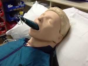
PIC1

(photo courtesy of Kai Romero, MD)

Why avoid endotracheal intubation?

Endotracheal intubation is a life-saving intervention when applied skillfully, but its attendant risks are well described.  Staving off intubation has been shown to decrease complications such as hypotension, arrhythmias, death, and nosocomial infections. Intubation also places patients at an increased need for sedation and invasive procedures2. Noninvasive ventilation has become commonplace in the ED to treat respiratory failure and prevent intubation1.

Some Definitions:

  • CPAP: applies constant pressure throughout the breathing cycle to increase functional residual capacity (FRC) by recruiting alveoli, decreasing work of breathing, and improving oxygenation. It is best given in hypoxemic patients 1,3.
  • PEEP/EPAP: Positive End Expiratory Pressure, which is the alveolar pressure before inspiratory flow begins. Adding PEEP helps decrease the amount of work required to initiate a breath. It also helps to decrease atelectasis1,4.
  • Bi-level: Cycled ventilation between Inspiratory Positive Airway Pressure (IPAP) and Expiratory Positive Airway Pressure/PEEP1BiPAP supports ventilation and increases oxygenation.
  • Pressure Support: The difference between EPAP and IPAP is referred to as pressure support. Pressure support makes it easier to draw larger tidal volumes1,4.

Applications for NPPV/Bi-level

1. Chronic Obstructive Pulmonary Disease (COPD)

In COPD, acute respiratory failure manifests as hypoxic, hypercapneic respiratory failure with collapse of small airways. Hyperinflation also occurs. Acute respiratory failure from COPD leads to increased work of breathing, acidosis, altered mental status, and ultimately coma, decompensation, and death5

Bi-level ventilation is a primary treatment option in COPD with good evidence for success5. When compared to usual medical care, bi-level ventilation decreases the risk of death (relative risk reduction 48%) and intubation rates (RRR 60%)5.

Number Needed to Treat (NNT) for mortality benefit = 10

NNT to prevent intubation = 4

Furthermore, when comparing patients with moderate and severe acidosis, bi-level ventilation decreased mortality, rates of intubation, and lengths of stay. It also improved work of breathing, acidosis, and PaC02 levels. Finally, regarding these outcomes, there were no significant differences between more and less acidotic patients at admission5,6.

Indications for NIPPV/bi-level ventilation6

1.      pH <7.35or PaC02 >45 mmHg

2.      Severe dyspnea with signs of increased work of breathing

3.      Caution: In severe respiratory acidosis (pH <7.25), failure rates of NIPPV may be as high as 50%1

Common Initial Settings:

  • IPAP 8-20 cm H2O (up to 30 cm H20)
  • EPAP 2-6 cm H2O to overcome intrinsic airway collapse3,5,7
  • Begin with either high IPAP and then titrate down, or low and titrate high. Both are reasonable, but require close monitoring to meet ventilation goals.7 Each patient is different.
  • Endpoints for physiologic improvement:  at 1 hour, reassessment should be made. Decisions regarding treatment failure, worsening clinical status of bi-level should be made early.5

2. Cardiogenic Pulmonary Edema (CPO)

Acute cardiogenic pulmonary edema is a common and potentially fatal cause of acute respiratory distress. CPO is related to a critical interaction between worsening left ventricular systolic function and an acute increase in systemic vascular resistance that results in rapid accumulation of fluid in the interstitium of the lung. This leads to decreased lung compliance, increased airway resistance, hypoxia, decreased diffusion capacity, and hypercarbia from muscle fatigue8.

Bi-level offers the advantage of improving both cardiac and pulmonary function by providing pressure support with IPAP and EPAP/PEEP. IPAP assists ventilation, which decreases the work of breathing and assures adequate ventilation. The EPAP/PEEP increases the FRC by recruiting collapsed alveoli, improving oxygenation, and helping to force interstitial fluid back into the pulmonary vasculature9,10.

Bi-level ventilation also increases intrathoracic pressure, which can lead to decreased left ventricular (LV) end diastolic volume. This results in decreased afterload and increased LV ejection fraction/stroke volume. Thus, the heart muscle is stretched less, and placed at a steeper part of the Starling curve. This results in stronger LV contractions, and reductions in BP and HR11.

PIC2

(Figure: in the failing heart, NIPPV both decreases preload and also shifts the Starling curve (decreases afterload) to augment cardiac performance)12,13

Common Initial Settings:

·         IPAP: 10 to 20 cm H20

·         EPAP: 5 to 10 cm H20

·         I:E ratio of IT to ET and is usually set at 1:3 or 1:4 (Inspiratory to Expiratory ratio)

Evidence for Bi-level ventilation in CPO:

Unfortunately, most of the evidence for NIPPV for CPO is centered on CPAP with few trials comparing the two modalities (CPAP and bi-level) head-to-head.

In a Cochrane review looking at NIPPV in CPO, CPAP alone has been proven to decrease intubation rates and to decrease in-hospital mortality, without the same benefit seen using bi-level ventilation14.

In the treatment of CPO, controversy regarding the safety of bi-level ventilation stems mainly from a single small study comparing the two NIPPV modalities. This study showed a more rapid improvement in vital signs, dyspnea and arterial blood gas (ABG) results with bi-level, but it also showed higher rates of myocardial infarction (MI) with bi-level ventilation15.  However, subsequent trials comparing CPAP and bi-level showed no difference in MI rates, but decreased intubation rates for those treated with bi-level, especially in patients presenting with hypercarbia16,17.

A trend that holds true is that bi-level leads to rapid improvement in physiological parameters such as respiratory rate, pH, PaCO2, PaO2, HR, work of breathing, afterload, preload, cardiac index, and ejection fraction. However,  more studies need to be performed to show a clear benefit in patient mortality and consistently decreased intubation rates18–21.

Indications for NIPPV in CPO:

1.      Increased work of breathing

2.      Hypercapnia and respiratory failure

3. Asthma

In asthma, NIPPV/bi-level ventilation might help to overcome intrinsic auto-PEEP, to decrease ventilation/perfusion (V/Q) mismatch by increased recruitment of alveoli, and to have a direct bronchodilatory effect in order to decrease work of breathing22.

In the mechanical ventilation of an asthmatic, one should carefully weigh the risk of critical error if the patient is not allowed a prolonged period of expiration in order to prevent “auto-PEEP”. Auto-PEEP is a condition where inhaled breaths are progressively delivered to lungs that have not returned to their FRC. This can subsequently lead to life-threatening hypotension and severe barotrauma, such as pneumothorax2.

Complications from intubation in a severe asthmatic can be severe, including cardiovascular collapse, pneumothorax, and prolonged intubation23.  However, a systematic review comparing NIPPV to medical care did not find a significant benefit to mortality or decreased rates of intubation, though these end points were likely limited by small numbers of patients studied in the intervention22.

Given the risks of intubation, and the purported physiologic benefits of bi-level ventilation to a severe asthmatic, NIPPV is a reasonable treatment strategy.  However, this should be done in tandem with medical management in carefully selected and monitored patients. Of note, NIPPV in asthma has not yet achieved the standard of care.

Indications for NIPPV in Asthma:

1.      As a carefully applied adjunct to medical management in refractory asthma

4. Acute Respiratory failure (no pre-existing chronic lung disease)

The benefits of NIPPV is not clear in undifferentiated acute respiratory failure and the evidence is often conflicting.  The use of NIPPV in undifferentiated acute respiratory failure (ARF) has shown similar mortality rates to conventional ventilation, but it has also demonstrated decreased intubation rates. On the other hand, decreased intubation rates may not apply to those with pneumonia or non-hypercapneic respiratory failure (PNA)24.

A recent multi-center trial showed no decrease in intubation rates for non-hypercapneic ARF (64% PNA) in those without chronic lung disease when compared to high flow or regular oxygen. Additionally, 90 day mortality was decreased in the high flow oxygen group compared to NIPPV. Also, delayed intubation via NIPPV did not show an association with increased mortality25.

Method of delivery and severity of illness likely affect mortality rates in patients with adult respiratory distress syndrome (ARDS) treated with NPPV, as a recent RCT showed decreased 90 day mortality in patients ventilated with helmeted NPPV compared to face mask NPPV26.

Indications:

1.      There are no clearly recommended indications and it should be on a case by case basis.

5. Blunt Thoracic Trauma

In a recent meta-analysis of 219 patients with thoracic trauma, NIPPV decreased intubation rates, improved oxygenation, decreased infection rates and showed mortality rates of 3% vs 22.9% in “standard management,” which was CPAP or face mask oxygen27.

6. Special Populations:

NIPPV decreases intubation rates and in-hospital mortality when applied to acute hypoxic respiratory failure in immunocompromised patients1,28.

It may also provide a benefit to patients with palliative care needs in whom endotracheal intubation is not within their goals of care, with some studies showing reversal of acute respiratory failure and return to home1,28,29.

The use of NPPV in the ARF of decompensation of neuromuscular disease is controversial, and may not be indicated in those with rapidly progressive disease29.

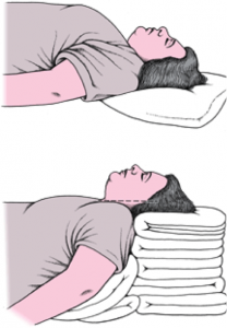
Pre-oxygenation For Intubation:

Much of the focus has been on avoiding intubation, but in critically ill patients, intubation and mechanical ventilation can be life-saving; to intubate an unstable patient is perilous, with desaturation, arrhythmia, cardiovascular collapse and cardiac arrest as well recognized entities30.

Pre-oxygenation prior to intubation is the standard of care to prevent life threatening complications during intubation.  Some patients display refractory hypoxia in the face of usual facemask pre-oxygenation, and failure to reach 93-95% Sp02 prior to intubation places the patient at risk for desaturation and apnea during the procedure31.

NIPPV has been shown to decrease desaturation rates in refractory hypoxia during pre-oxygenation for intubation and should be strongly considered for pre-oxygenation prior to intubation in refractory hypoxia30–32.

Pearls:

  1. Positive Pressure is not everything. Do not forget medical management
  2. Bi-level ventilation is an early “go-to” in moderate to severe COPD exacerbations
  3. Experienced respiratory therapists (RTs) and staff are critical to the success of non-invasive ventilation
  4. Once applied, reassess your patient frequently and be ready to adjust!
  5. Can be used to stave off intubation at the end of life
  6. Consider for pre-oxygenation prior to intubation

Pitfalls:

  1. Positive pressure can cause hypotension and decompensation if blindly applied
  2. Do not place a pressure mask on a damaged face or a fluid filled mouth
  3. Do not delay necessary intubation
  4. Do not let your patient Auto-PEEP
  5. Your severely acidotic patient is at high risk for failure: be ready to intubate.
  6. If your patient is not awake, then the patient should be intubated.

 Who Needs It?

  1. Patients with moderate to severe COPD exacerbations
  2. Those patients with cardiogenic pulmonary edema with increased work of breathing or hypercapnia
  3. Patients with isolated blunt thoracic trauma
  4. The immunocompromised patient with hypoxic respiratory failure
  5. Patients who require pre-oxygenation prior to intubation

Who Does Not?

  1. Altered Mental Status
  2. Facial Trauma/Can’t Handle their own secretions

Gray Zones?

  1. Asthma
  2. Neuromuscular Disease
  3. Undifferentiated Hypoxic Respiratory Failure

References / Further Reading

1. Aboussouan, L. S. & Ricaurte, B. Noninvasive positive pressure ventilation: Increasing use in acute care. Cleve. Clin. J. Med. 77, 307–316 (2010).

2. Leatherman, J. Mechanical ventilation for severe asthma. Chest 147, 1671–1680 (2015).

3. Bolton, R. & Bleetman, A. Non-invasive ventilation and continuous positive pressure ventilation in emergency departments: where are we now? Emerg. Med. J. EMJ 25, 190–194 (2008).

4. Appendini, L. et al. Physiologic effects of positive end-expiratory pressure and mask pressure support during exacerbations of chronic obstructive pulmonary disease. Am. J. Respir. Crit. Care Med. 149, 1069–1076 (1994).

5. Ram, F. S. F., Picot, J., Lightowler, J. & Wedzicha, J. A. Non-invasive positive pressure ventilation for treatment of respiratory failure due to exacerbations of chronic obstructive pulmonary disease. Cochrane Database Syst. Rev. CD004104 (2004). doi:10.1002/14651858.CD004104.pub2

6. Pauwels, R. A., Buist, A. S., Calverley, P. M. A., Jenkins, C. R. & Hurd, S. S. Global Strategy for the Diagnosis, Management, and  Prevention of Chronic Obstructive Pulmonary Disease. Am. J. Respir. Crit. Care Med. 163, 1256–1276 (2001).

7. Prinianakis, G., Delmastro, M., Carlucci, A., Ceriana, P. & Nava, S. Effect of varying the pressurisation rate during noninvasive pressure support ventilation. Eur. Respir. J. 23, 314–320 (2004).

8. Wiesen, J., Ornstein, M., Tonelli, A. R., Menon, V. & Ashton, R. W. State of the evidence: mechanical ventilation with PEEP in patients with cardiogenic shock. Heart Br. Card. Soc. 99, 1812–1817 (2013).

9. Cross, A. M. Review of the role of non-invasive ventilation in the emergency department. J. Accid. Emerg. Med. 17, 79–85 (2000).

10. Peter, J. V., Moran, J. L., Phillips-Hughes, J., Graham, P. & Bersten, A. D. Effect of non-invasive positive pressure ventilation (NIPPV) on mortality in patients with acute cardiogenic pulmonary oedema: a meta-analysis. Lancet Lond. Engl. 367, 1155–1163 (2006).

11. Sheldon, R. Congestive heart failure and noninvasive positive pressure ventilation. Emerg. Med. Serv. 34, 64–67 (2005).

12. Figueroa, M. S. & Peters, J. I. Congestive heart failure: Diagnosis, pathophysiology, therapy, and implications for respiratory care. Respir. Care 51, 403–412 (2006).

13. Shekerdemian, L. & Bohn, D. Cardiovascular effects of mechanical ventilation. Arch. Dis. Child. 80, 475–480 (1999).

14. Vital, F. M. R., Ladeira, M. T. & Atallah, A. N. Non-invasive positive pressure ventilation (CPAP or bilevel NPPV) for cardiogenic pulmonary oedema. Cochrane Database Syst. Rev. CD005351 (2013). doi:10.1002/14651858.CD005351.pub3

15. Mehta, S. et al. Randomized, prospective trial of bilevel versus continuous positive airway pressure in acute pulmonary edema. Crit. Care Med. 25, 620–628 (1997).

16. Nava, S. et al. Noninvasive ventilation in cardiogenic pulmonary edema: a multicenter randomized trial. Am. J. Respir. Crit. Care Med. 168, 1432–1437 (2003).

17. Gray, A. J. et al. A multicentre randomised controlled trial of the use of continuous positive airway pressure and non-invasive positive pressure ventilation in the early treatment of patients presenting to the emergency department with severe acute cardiogenic pulmonary oedema: the 3CPO trial. Health Technol. Assess. Winch. Engl. 13, 1–106 (2009).

18. Park, M. et al. Oxygen therapy, continuous positive airway pressure, or noninvasive bilevel positive pressure ventilation in the treatment of acute cardiogenic pulmonary edema. Arq. Bras. Cardiol. 76, 221–230 (2001).

19. Crane, S. D., Elliott, M. W., Gilligan, P., Richards, K. & Gray, A. J. Randomised controlled comparison of continuous positive airways pressure, bilevel non-invasive ventilation, and standard treatment in emergency department patients with acute cardiogenic pulmonary oedema. Emerg. Med. J. EMJ 21, 155–161 (2004).

20. Levitt, M. A. A prospective, randomized trial of BiPAP in severe acute congestive heart failure. J. Emerg. Med. 21, 363–369 (2001).

21. Pang, D., Keenan, S. P., Cook, D. J. & Sibbald, W. J. The effect of positive pressure airway support on mortality and the need for intubation in cardiogenic pulmonary edema: a systematic review. Chest 114, 1185–1192 (1998).

22. Ram, F. S. F., Wellington, S., Rowe, B. & Wedzicha, J. A. Non-invasive positive pressure ventilation for treatment of respiratory failure due to severe acute exacerbations of asthma. Cochrane Database Syst. Rev. CD004360 (2005). doi:10.1002/14651858.CD004360.pub3

23. Landry, A., Foran, M. & Koyfman, A. Does Noninvasive Positive-Pressure Ventilation Improve Outcomes in Severe Asthma Exacerbations? Ann. Emerg. Med. 62, 594–596

24. Honrubia, T. et al. Noninvasive vs conventional mechanical ventilation in acute respiratory failure : A multicenter, randomized controlled trial. Chest 128, 3916–3924 (2005).

25. Frat, J.P. et al. High-flow oxygen through nasal cannula in acute hypoxemic respiratory failure. N. Engl. J. Med. 372, 2185–2196 (2015).

26. Patel, B. K., Wolfe, K. S., Pohlman, A. S., Hall, J. B. & Kress, J. P. Effect of Noninvasive Ventilation Delivered by Helmet vs Face Mask on the Rate of Endotracheal Intubation in Patients With Acute Respiratory Distress Syndrome: A Randomized Clinical Trial. JAMA 315, 2435–2441 (2016).

27. Chiumello, D., Coppola, S., Froio, S., Gregoretti, C. & Consonni, D. Noninvasive ventilation in chest trauma: systematic review and meta-analysis. Intensive Care Med. 39, 1171–1180 (2013).

28. Hill, N. S., Brennan, J., Garpestad, E. & Nava, S. Noninvasive ventilation in acute respiratory failure. Crit. Care Med. 35, 2402–2407 (2007).

29. Mas, A. & Masip, J. Noninvasive ventilation in acute respiratory failure. Int. J. Chron. Obstruct. Pulmon. Dis. 9, 837–852 (2014).

30. Mosier, J. M. et al. The Physiologically Difficult Airway. West. J. Emerg. Med. 16, 1109–1117 (2015).

31. Weingart, S. D. & Levitan, R. M. Preoxygenation and prevention of desaturation during emergency airway management. Ann. Emerg. Med. 59, 165–175.e1 (2012).

32. Baillard, C. et al. Noninvasive ventilation improves preoxygenation before intubation of hypoxic patients. Am. J. Respir. Crit. Care Med. 174, 171–177 (2006).


The post Bi-level Ventilation: Who Needs it and Who Doesn’t? Pearls and Pitfalls appeared first on emdocs.

R.E.B.E.L. EM – Is Apneic Oxygenation Overhyped? with Scott Weingart

$
0
0

Originally published at R.E.B.E.L. EM on April 4, 2016. Reposted with permission.

Follow Dr. Salim R. Rezaie (@srrezaie) and Dr. Scott Weingart (@emcrit

Apneic-Oxygenation-765x583Welcome back to the April 2016 edition of REBELCast. For this episode I was lucky enough to get Scott Weingart on the show to talk to us about all things Apneic Oxygenation (ApOx). ApOx is a concept that has been around for some time in the operating room literature, but only recently been gaining acceptance in the ED, especially after the publication of this concept by Scott and Richard Levitan in the Annals of Emergency Medicine in 2011 [1]. Many nay sayers will argue that the OR studies were in controlled settings with elective surgical patients who were not in critical condition. The believers would argue that ApOx makes sense, its low cost,  and low complexity.  To date there has been no randomized controlled trials (RCTs) on ApOx in the ED.  There has been one ICU Trial (i.e. The FELLOW Trial) [2] and an even more recent observational trial in the ED [3] that have been published on the topic of ApOx. So the question remains: Is Apneic Oxygenation Overhyped?

What are Preoxygenation (PreOx), Apneic Oxygenation (ApOx), and Reoxygenation (ReOx)?

Preoxygenation (PreOx)

  • Should be broken up into 2 separate terms: Preoxygenation and Denitrogenation
  • Denitrogenation = Washing out as much nitrogen from the lungs so that you have a buffer/bag of oxygen when the patient isn’t breathing
  • Requires Time: 3 min of tidal volume breathing on a high FiO2 source
    • With a Non-rebreather (NRB) mask alone,  you are giving approximately 60% FiO2 which will make it impossible to accomplish denitrogenation
  • Preoxygenation (PreOx) = Getting the O2 saturation as close to 100% before pushing RSI meds to intubate

Apneic Oxygenation (ApOx)

  • This occurs during the time from pushing intubation medications, which is anywhere from 45 – 60 seconds, while the paralytic is taking effect, that the patient is burning through their oxygen stores
  • Apneic Oxygenation (ApOx) = Passive movement of oxygen to the alveoli without the patient having to breath and without you having to breath for them
  • Apneic CPAP = Little Brother of ApOx; In patients who have closed alveoli or flooded alveoli; Essentially used for maintenance of recruitment of alveoli during apneic period

Apneic CPAP

Reoxygenation (ReOx)

  • Reoxygenation (ReOx) = Attempts to increase O2 sats when a patient drops their sats during airway management
  • If patient has physiologic shunt physiology, standard BVM will not suffice to fix patients desaturation in between attempts at intubation

BOTTOM LINE: PreOx, ApOx, and ReOx are all attempts to avoid the deadly DeOx (Deoxygenation)

What are your thoughts on the The Fellow Trial? [2]

  • Summary of Trial:
    • Randomized Controlled Trial (RCT) of 150 Critically Ill Patients in a Single ICU
    • Randomized to Apneic Oxygenation vs Usual Care
    • Study Conclusion: Use of Apneic Oxygenation vs Usual Care Made no Difference in the Lowest Arterial Oxygen Saturation Between Induction and Two Minutes After Completion of Intubation
  • Issue with Usual Care in this Trial:
    • Majority of patients had BVM during apneic period
      • Most ED patients are not fasted and using BVM could potentially cause vomiting
    • Not maintaining an open pathway from the nares to the glottis for patients not being bagged
    • Combination of these 2 things hurt the take home message of the study

Does Apneic Oxygenation help in patients with pulmonary shunt physiology (i.e. pulmonary edema, multifocal pneumonia, etc…)?

  • ApOx will help if it is being given with Apneic CPAP
  • Without CPAP, ApOx will not work in patients with shunt physiology
  • Bagging with BVM alone will give O2 and PEEP but again we really want to avoid bagging our patients in the ED as this can cause vomiting
    • With this strategy ApOx may be superfluous
  • A better strategy might be NC at 15 LPM + BVM (without bagging) and with a PEEP Valve

What are your thoughts on the Observational ED Trial recently published by Sackles et al? [3]

  • Summary of Trial:
    • Observational trial in a single ED of 635 patients who received either ApOx or No ApOx
    • Study Conclusion: ApOx in Adult Patients Undergoing RSI had better 1st pass intubation without hypoxemia with a NNT of 7.6
  • The Fact that this Trial is Observational:
    • For People Who Believe in ApOx: Helpful trial that confirms their belief structure
    • For People Who Don’t Believe in ApOx: Does not change their minds, because this is still only observational data
  • Bottom Line:  We need an ED RCT on ApOx

Can you walk us through your exact approach to preoxygenation in a septic patient with pneumonia who is tachypneic, hypoxic, and hypotensive?

  • The Physiologically Difficult Airway (HOp Killers)
    • Hypotension
    • Oxygenation (i.e. Hypoxemia)
    • pH and Ventilation
  • How to Manage our Patient with Hypotension and Hypoxemia
    • Oxygenation (i.e. Hypoxemia)
      • Place patient on standard nasal cannula (NC) at 15L
      • BVM with PEEP valve with 2 hand mask seal for 3 minutes (NO NEED TO BAG) for  preoxygenation and denitrogenation
    •  Hypotension
      • RSI Meds (Great Explanation HERE)
        • Ketamine IV 0.5 mg/kg
        • Rocuronium IV 1.6 – 2.0 mg/kg
      • Start norepinephrine drip or push aliquots of push-dose epinephrine
        • Norepinephrine IV 0.01 – 1 mcg/kg/min
        • Push-Dose Epinephrine IV 5 – 20 mcg every 2 – 5 min

Push-Dose Pressor

Image Borrowed from emcrit.org

Is there a patient we should not use ApOx in?

  • We know ApOx Works in Patients Without Shunt Physiology
    • THRIVE Trial: Transnasal humidified Rapid-Insufflation Ventilatory Exchange [4]
      • 25 patients with difficult airways undergoing general anesthesia
      • Median Apnea was 14 minutes
      • No patient experienced O2 Sat <90%
  • Most Modalities we use for Pre-Oxygenation and Denitrogenation are NOT good enough on their own (i.e. Non-Rebreather Mask)
  • BOTTOM LINE: There is no reason at this time to not be using nasal cannula for ApOx with intubation

Take Home Messages:

  • PreOx is getting the O2 saturation as close to 100% before pushing RSI meds to intubate
  • ApOx is passive movement of oxygen to the alveoli without the patient having to breath and without you having to breath for them
  • In patients with pulmonary shunt physiology, a better strategy might be NC at 15LPM + BVM (Without Bagging) + a PEEP Valve because this will provide both O2 and CPAP for alveolar recruitment
  • The Physiologically Difficult Airway = HOp Killers
    • Hypotension
    • Oxygenation (i.e. Hypoxemia)
    • pH and Ventilation
  • There is no reason at this time to not be using nasal cannula for ApOx with intubation

BONUS Question:

Lets say for a moment that you are not Scott Weingart.  You are working as a faculty at a teaching institution and you have a resident approach you saying they had just listened to the EMCrit podcast. They want to try something because they heard it on the podcast.  How would you handle that situation?

  • Things discussed on the EMCrit podcast are things that are able to be done in the environment of an ED ICU with fellows, which is different than what most people have.
  • In general, as a resident you shouldn’t say you heard something on a podcast and want to do it:
    • Puts people in a tough situation
    • Its important to read the primary literature behind what is being said
  • As the attending who is hearing this, a good way to handle this is telling the resident:
    • Lets discuss this more after we get our patient stabilized
    • Lets do a journal club on this and see if this is something we can incorporate into our practice

For More on This Topic Checkout:

References:

  1. Weingart, Scott D, and Richard M Levitan. Preoxygenation and prevention of desaturation during emergency airway management. Ann Emerg Med 2011; 59 (3): 165 – 75. PMID: 22050948
  2. Semler MW et al. Randomized Trial of Apneic Oxygenation During Endotracheal Intubation of the Critically Ill. Am J Respir Crit Care Med 2015 [Epub ahead of print] PMID: 26426458
  3. Sackles JC et al. First Pass Success Without Hypoxemia is Increased with the Use of Apneic Oxygenation During RSI in the Emergency Department. Acad Emerg Med 2016. [epub ahead of print] PMID: 26836712
  4. Patel A et al. Transnasal Humidified Rapid-Insufflation Ventilatory Exchange (THRIVE): a physiological method of increasing apnoea time in patients with difficult airways. Anesthesia 2015; 70: 323 – 329. PMID: 25388828

Post Peer Reviewed By: Anand Swaminathan (Twitter: @EMSwami)

The post R.E.B.E.L. EM – Is Apneic Oxygenation Overhyped? with Scott Weingart appeared first on emdocs.

Pearls for the management of GSW associated traumatic injury

$
0
0

Author: Joshua Bucher, MD (Assistant Professor, Department of EM, Rutgers – RWJMS; Assistant EMS Medical Director, RWJ-MHS) // Edited by: Alex Koyfman, MD (@EMHighAK, EM Attending Physician, UTSW / Parkland Memorial Hospital) and Brit Long, MD (@long_brit, EM Attending Physician at SAUSHEC)

Case

A 27-year-old male is brought in by EMS after sustaining several gunshot wounds. On arrival, the patient withdraws from painful stimuli, does not open his eyes and makes incomprehensible noises. His heart rate is 130 bpm, blood pressure 84/42, RR 26, and Sp02 88%. While preparing to begin resuscitation, what are the first and most important steps?

Airway

As with all critical patients, the first step is airway management. Care should be taken and best practices should be followed to allow for first pass success including preoxygenation if possible to avoid desaturation. Resuscitation before intubation if possible is important. The appropriate induction agent is of vital importance. Ketamine is associated with the most neutral hemodynamic properties, and it is also the ideal agent for head injured patients.1,2 By maintaining hemodynamic stability and its dissociative properties, it is useful to blunt the response to laryngoscopy. Fentanyl is another option as well at doses 3-5 mcg/kg IV.3

Gunshot wounds may directly involve airway structures. In that case, surgical airway may be preferred, compared to endotracheal intubation. Team preparation is key for this step, and verbalizing the need for potential surgical airway is essential.

C-Spine

Along with airway management, unless there is a focal neurologic injury, it is not necessary to perform cervical spine immobilization of patients suffering from gunshot wounds. This is based on a strong retrospective study as well as supported by the NAEMSP/ACS-COT position paper on spinal immobilization.4 Cervical spine immobilization can directly interfere with airway management, obscure the mouth opening, require increased laryngoscopic force and can lead to worse patient outcomes in the setting of a failed airway.5-7

Breathing

Breathing can be significantly affected depending on where your patient has been shot. Any sign of a tension pneumothorax (decreased breath sounds on one side, tracheal deviation, or hemodynamic instability) needs to be immediately treated with needle decompression or finger thoracostomy followed by tube thoracostomy. A standard pneumothorax can be treated with tube thoracostomy, either during the primary survey or after. High flow NRB oxygen is indicated as well. A GSW to the abdomen may cause difficulty with ventilation due to pain or abdominal distention, and this needs to be monitored.

There have been some newer developments in the management of pneumo/hemothoraces. Inaba et al. described their experience using a smaller chest tube catheter for traumatic pneumo- or hemothoraces and found no difference in patient outcomes with 28-32 vs traditional 36-40 French chest tubes.8 Furthermore, Russo et al. studied a new method of using a pigtail catheter in a swine model vs traditional chest tube and found that both were able to drain the same amount of blood from a hemothorax.9 Although pigtail catheters have been used for pneumothoraces, this is a good step forward towards utilization of more patient oriented resources in the management of a hemothorax. The EAST guidelines currently recommend tube thoracostomy for all hemothoraces. They also suggest that occult pneumothoraces be treated with observation in a stable patient, even with positive pressure ventilation.10

Circulation

The next step of the primary survey is circulation. At this point, fluid resuscitation should begin with blood products in the critically injured patient. Permissive hypotension should be considered to target a systolic of 90 mm Hg or a MAP or 45 – 50 mm Hg, although further prospective studies are required.11  In addition, blood products should be transfused in a 1:1:1 ratio of 6 units of PRBCs:6 units FFP:1 pack of platelets, based on the results of the PROPPR trial.12 Furthermore, crystalloid fluids should be limited unless absolutely necessary to maintain perfusion.13 Fluids can theoretically prohibit clotting and dilute hemoglobin carrying capacity, and this recommendation is supported by the EAST guidelines.14

Thoracotomy

Emergency department thoracotomy is a life-saving procedure for patients with a very low survival. The EAST guidelines define signs of life as pupillary response, ventilation, vital signs, cardiac electrical activity, or extremity movement. They released the following evidence-based recommendations.

screen-shot-2016-09-11-at-4-36-18-am

Recently, there has been research looking at this issue. Inaba et al prospectively studied patients undergoing resuscitative thoracotomy in the ER and related it to the FAST exam. They found that if the FAST exam was negative for pericardial fluid or any cardiac activity, the sensitivity was 100% for predicting the patient would not survive.15  This can be added to the EAST guidelines to determine the efficacy and necessity of thoracotomy.

REBOA

Resuscitative endovascular balloon occlusion of the aorta (REBOA) is a last-ditch procedure that can stop hemorrhagic shock in patients with bleeding below the diaphragm. The instrument is comprised of a sheath and balloon, which is inserted into the femoral artery and inflated at one of three areas in order to stop blood flow. This can be especially useful for pelvic fractures with hemorrhage or other intra-abdominal hemorrhagic processes. There currently is limited data since it is a novel device, but it appears to be promising for specific situations. You can read more about REBOA at http://lifeinthefastlane.com/ccc/resuscitative-endovascular-balloon-occlusion-aorta-reboa/.

Extremity Hemorrhage

I want to briefly mention two specific interventions that are geared towards pre-hospital providers. The first intervention is the use of tourniquets for extremity trauma. We now have a large body of literature that supports the use of tourniquets as a life-saving device for extremity trauma with minimal risk of side effects.16 Likewise, the use of clotting agents, such as the commercially named QuickClot agent, are safe and effective to stop bleeding and are recommended by the Tactical Combat Casualty Care guidelines for hemorrhage not amenable to tourniquet placement.16 These two options are highly efficacious and warrant our attention.

Case resolution:

The patient is intubated appropriately. Bilateral chest tubes are placed, with immediate return of 2L from the left chest and a large rush of air from the right. Massive transfusion protocol is activated, and the patient is immediately transfused blood and plasma products. An E-FAST is performed, showing no pericardial fluid but large intraabdominal fluid. The patient is taken to the operating room by the trauma team with successful repair of his thoracic and abdominal injuries and makes a full recovery.


Take Home Points

  1. Utilize ketamine for airway management as it is the most hemodynamically neutral agent.
  2. Use traditional large chest tubes for hemothoraces and large pneumothoraces.
  3. Aggressively resuscitate with blood products for the exsanguinating trauma patient.


References / Further Reading

  1. Bucher J, Koyfman A. Intubation of the Neurologically Injured Patient. The Journal of emergency medicine. 2015;49(6):920-927.
  2. Cohen L, Athaide V, Wickham ME, Doyle-Waters MM, Rose NG, Hohl CM. The Effect of Ketamine on Intracranial and Cerebral Perfusion Pressure and Health Outcomes: A Systematic Review. Annals of emergency medicine. 2014.
  3. Pouraghaei M, Moharamzadeh P, Soleimanpour H, et al. Comparison between the effects of alfentanil, fentanyl and sufentanil on hemodynamic indices during rapid sequence intubation in the emergency department. Anesthesiology and pain medicine. 2014;4(1):e14618.
  4. White Iv CC, Domeier RM, Millin MG, Standards, Clinical Practice Committee NAoEMSP. EMS Spinal Precautions and the Use of the Long Backboard -Resource Document to the Position Statement of the National Association of EMS Physicians and the American College of Surgeons Committee on Trauma. Prehospital emergency care : official journal of the National Association of EMS Physicians and the National Association of State EMS Directors. 2014;18(2):306-314.
  5. Gruen RL, Jurkovich GJ, McIntyre LK, Foy HM, Maier RV. Patterns of errors contributing to trauma mortality: lessons learned from 2,594 deaths. Annals of surgery. 2006;244(3):371-380.
  6. Santoni BG, Hindman BJ, Puttlitz CM, et al. Manual in-line stabilization increases pressures applied by the laryngoscope blade during direct laryngoscopy and orotracheal intubation. Anesthesiology. 2009;110(1):24-31.
  7. Goutcher CM, Lochhead V. Reduction in mouth opening with semi-rigid cervical collars. British journal of anaesthesia. 2005;95(3):344-348.
  8. Inaba K, Lustenberger T, Recinos G, et al. Does size matter? A prospective analysis of 28-32 versus 36-40 French chest tube size in trauma. The journal of trauma and acute care surgery. 2012;72(2):422-427.
  9. Russo RM, Zakaluzny SA, Neff LP, et al. A pilot study of chest tube versus pigtail catheter drainage of acute hemothorax in swine. The journal of trauma and acute care surgery. 2015;79(6):1038-1043; discussion 1043.
  10. Mowery NT, Gunter OL, Collier BR, et al. Practice management guidelines for management of hemothorax and occult pneumothorax. The Journal of trauma. 2011;70(2):510-518.
  11. Dunser MW, Takala J, Brunauer A, Bakker J. Re-thinking resuscitation: leaving blood pressure cosmetics behind and moving forward to permissive hypotension and a tissue perfusion-based approach. Critical care. 2013;17(5):326.
  12. Holcomb JB, Tilley BC, Baraniuk S, et al. Transfusion of plasma, platelets, and red blood cells in a 1:1:1 vs a 1:1:2 ratio and mortality in patients with severe trauma: the PROPPR randomized clinical trial. JAMA : the journal of the American Medical Association. 2015;313(5):471-482.
  13. Chatrath V, Khetarpal R, Ahuja J. Fluid management in patients with trauma: Restrictive versus liberal approach. Journal of anaesthesiology, clinical pharmacology. 2015;31(3):308-316.
  14. Cotton BA, Jerome R, Collier BR, et al. Guidelines for prehospital fluid resuscitation in the injured patient. The Journal of trauma. 2009;67(2):389-402.
  15. Inaba K, Chouliaras K, Zakaluzny S, et al. FAST ultrasound examination as a predictor of outcomes after resuscitative thoracotomy: a prospective evaluation. Annals of surgery. 2015;262(3):512-518; discussion 516-518.
  16. Defense Do. Tactical Combat Casualty Care Guidelines for Medical Personnel 2015.


The post Pearls for the management of GSW associated traumatic injury appeared first on emdocs.

Can’t Intubate Can’t Ventilate

$
0
0

Originally published at Pediatric EM Morsels on May 20, 2016. Reposted with permission.

Follow Dr. Sean M. Fox on twitter @PedEMMorsels

transtracheal-ventilation-attachments

“Can’t Intubate Can’t Ventilate” is one of the frightening statements that causes massive surges of adrenaline in everyone. Unfortunately, most neural synapses don’t function well with that large surge of adrenaline, and it is, therefore, imperative to contemplate how to manage this scenario before it arises.  We have previously discussed Transtracheal Ventilation and have several videos to view, but let us review this important topic briefly once more. Can’t Intubate Can’t Ventilate: How Do I Oxygenate?


Can’t Intubate Can’t Ventilate: Anatomy Matters!

  • With larger children and adults, the can’t intubate can’t ventilate scenario often leads to the Cricothyrotomy.
  • In younger children and infants, the differences in anatomy make a traditional cricothyrotomy challenging.
  • In infants and young children:
    • Generous proportions of subcutaneous adipose tissue (chunky little babies are cute…) obscures landmarks.
    • The Hyoid bone is more prominent than the thyroid cartilage.
    • The Thyroid notch is often not palpable.
    • The Cricothyroid membrane is:
      • More horizontally positioned vs its typical vertical position
      • Small!
        • Around 8 years of age it is 1/2 the height and width of an adult’s
        • In neonates, the size is not sufficient enough to insert any commonly used rescue device. [Navsa, 2005]
  • The altered anatomy makes location of the cricothyroid membrane more difficult (if at all possible) and the small size may make it impossible to pass a large cric-tube through.


Can’t Intubate Can’t Ventilate: Go Transtracheal

  • This is THE MOST IMPORTANT PROCEDURE TO KNOW!
  • Transtracheal ventilation has been used successfully in children as well as adults. [Frerk, 2015; Cote, 2009]
  • It may not “secure” an airway, but it will provide the patient with oxygen while you sort out the problem (and change your pants).
  • It is also easier than placing an IV in a child!
    • Locate the trachea!
      • If you are able to locate the cricothyroid membrane and it is large enough you can use it
      • Potential to use this catheter later to convert to a guidewire-assisted percutaneous cricothyrotomy. [Boccio, 2015]
    • Load a large gauge needle/catheter (14 gauge), ideally one that is reinforced(as simple peripheral IV catheters are prone to kink and become obstructed) onto a fluid-filled syringe.
    • Aspirate as you enter the skin at a 30-45 degree angle aimed caudally.
    • When you aspirate bubbles, you are in the airway! Advance the catheter and retract the needle.
    • Boom… done. High-Fives all around! {oh wait… we need oxygen!}


Can’t Intubate Can’t Ventilate: The Hard Part

  • The most difficult aspect of the procedure is not waiting too long to do it and leading to hypoxic insult.
  • The next most difficult aspect is figuring out how to connect oxygen to the tiny catheter you just placed in the neck.
  • This is where contemplation of how to do this before you need to do it is important, because most of us are not going to successfully “MacGyver it” on the fly.
  • Oxygen Connection Options

    1. Commercial products
      • Have flow regulators that are easy to use. [Cote, 2009]
      • Connect easily via Lure-lock to the catheter.
      • Many have pressure regulators as well.
      • Con = Expensive.
    2. Oxygen Tubing and High Flow O2 from Wall 
      • Not as optimal as commercial products, but may be best you have available.
      • Turn flow up all of the way. [Bould, 2008]
      • Need to “MacGyver” a flow regulator and a connector
        • Flow Regulator
          • Cut large holes (several) in side of oxygen tubing.
          • Need large/multiple holes to allow air flow to egress easily and not add to PEEP. [Sasano, 2014]
          • May also use Y-connector to another oxygen tube.
        • Connector
          • 3-way stop cock can be used to fit into distal end of oxygen tubing and Lure-lock onto the catheter.
          • Need to ensure 3 way valve is open to flow!
    3. Self-Inflating Ventilation Bag [Sasano, 2014]
      • Not as optimal as commercial products, but may be best you have available.
      • 3.0 ETT bag connector
        • Remove from ETT
        • Insert distal end into catheter
      • 7.5 ETT bag connector
        • Remove from ETT
        • Insert into proximal end of 3 mL syringe (after removing the plunger).
        • Use Lure-lock on syringe to connect to catheter
      • Will need to disengage the bag’s pop-off valve.
  • Oxygenate!
    • Occluding the flow regulator will lead to airflow into the trachea (inspiration).
    • Uncovering the flow regulator will allow air flow from oxygen source and patient to escape (expiration).
    • Inspiration : Expiration = 1 second :  4 seconds
    • Use longer expiration phases for completely occluded upper airway (ex, 1:9)
      • Patient will tolerate hypercapnia better than barotrauma/pneumothorax.


Moral of the Morsel

  • Do not let the first time you think about transtracheal ventilation be when you realize you need to do it.
  • Know what equipment you have available.
    • If you have a commercial product, know how to use it and where it is.
    • If you don’t have a commercial product, make your MacGyver survival bag and keep it handy with the tools you need, so you don’t need to recall how to do it in the time of need.


References

Boccio E1, Gujral R2, Cassara M3, Amato T4, Wie B5, Ward MF6, D’Amore J7. Combining transtracheal catheter oxygenation and needle-based Seldinger cricothyrotomy into a single, sequential procedure. Am J Emerg Med. 2015 May;33(5):708-12. PMID: 25791154. [PubMed] [Read by QxMD]
Frerk C1, Mitchell VS2, McNarry AF3, Mendonca C4, Bhagrath R5, Patel A6, O’Sullivan EP7, Woodall NM8, Ahmad I9; Difficult Airway Society intubation guidelines working group. Difficult Airway Society 2015 guidelines for management of unanticipated difficult intubation in adults. Br J Anaesth. 2015 Dec;115(6):827-48. PMID: 26556848. [PubMed] [Read by QxMD]
Bould MD1, Bearfield P. Techniques for emergency ventilation through a needle cricothyroidotomy. Anaesthesia. 2008 May;63(5):535-9. PMID: 18412654. [PubMed][Read by QxMD]

The post Can’t Intubate Can’t Ventilate appeared first on emdocs.

R.E.B.E.L. EM – Critical Care Updates: Resuscitation Sequence Intubation – Hypoxemia Kills (Part 2 of 3)

$
0
0

Originally published at R.E.B.E.L. EM on September 29, 2017. Reposted with permission.

Follow Dr. Salim R. Rezaie at @srrezaie 


This blog post is the second part of a series of 3, on a recent lecture I was asked to give  on Critical Care Updates: Resuscitation Sequence Intubation. This talk was mostly derived from a podcast by Scott Weingart (Twitter: @EMCrit) where he talked about the physiologic killers during preintubation and perintubation. In this podcast, Scott mentions the HOp killers: Hypotension, Hypoxemia, and Metabolic Acidosis (pH) as the physiologic causes of pre-intubation/peri-intubation morbidity and mortality. Taking care of these critically ill patients that require intubation can be a high stress situation, with little room for error.  In part two of this series we will discuss some useful strategies at the bedside to help us reduce pre-intubation/peri-intubation hypoxemia.

What was the Premise of this Talk?

  • Resuscitate Before You Intubate
  • Hefner AC et al [1] and Kim WY et al [2] evaluated over 2800 patients requiring emergency intubation. In both trials the rate of cardiac arrest (CA) within 10 minutes of intubation ranged from 1.7% – 2.4%. Both trials listed pre-intubation hypotension (SBP ≤90mmHg) as a risk factor for cardiac arrest. Hefner AC et al also mentioned hypoxemia as a risk factor as well.

What are the Physiologic Killers Pre-intubation/Peri-intubation?

Hypoxemia Kills

  • The Basics:
    • Think NO DESAT (Nasal Oxygen During Efforts Securing A Tube)
    • NC at 15LPM + NRB at 15LPM (In all actuality you are turing the O2 all the way up, which may be more than 15LPM)
    • If you cannot get the O2 Saturation ≥95%, then consider the following:
      • Lung Shunt Physiology (i.e. Pulmonary Edema, Pneumonia, etc…).  These patients still need oxygen, but also need PEEP to recruit atelectatic alveoli to overcome the shunt
  • Intervention 1: NC 15LPM + BVM 15LPM + PEEP Valve 5 – 15cmH20
    • You don’t need to bag these patients, they need a tight seal and jaw thrust (i.e. Apneic CPAP Recruitment)
    • Bottom Line:  In critically ill patients in which you cannot get O2 Sats ≥95%, consider shunt physiology and use Apneic CPAP Recruitment
  • Intervention 2: Delayed Sequence Intubation (DSI)
    • Procedural sedation for the procedure of preoxygenation
      • Give 1mg/kg IV Ketamine -> Preoxygenate -> Paralyze the Patient -> Apneic Oxygenation -> Intubate
    • Evidence: Weingart et al Ann Emerg Med 2015 [1]
      • Observational trial with >150 intubations -> 62 patients uncooperative/combative
      • Mean O2 Saturation increased from 89.9% unto 98.8%
      • No Complications
    • Bottom Line:  In critically ill, agitated patients, who are hypoxemic, that need to be intubated, consider using DSI, which is procedural sedation for preoxygenation.
  • Intervention 3: Back Up Head Elevated (BUHE) Intubation [2]
    • 528 Intubations
    • Primary Outcome: Composite of Any Intubation-Related Complication (Difficult Intubation ≥3 Attempts or > 10 min, Hypoxemia <90% O2 Sat, Esophageal Intubation, or Esophageal Aspiration
    • Primary Outcome Results:
      • Standard Supine Intubation: 22.6%
      • BUHE Intubation 9.3%
    • Bottom Line: BUHE still needs prospective external validation in an ED setting, but seems to decrease intubation-related complications in comparison to standard supine intubation

Clinical Bottom Line:

  • Pre-Intubation Hypoxemia is a risk factor for Peri-Intubation Cardiac Arrest:
    Options to Improve Hypoxemia:
  • Start with NO DESAT (Nasal Oxygen During Efforts Securing A Tube) at 15LPM + NRB at 15LPM
  • If you don’t get the O2 Sats ≥ 95% think about shunt physiology and consider the following interventions:
    • Intervention 1: NC 15LPM + BVM 15LPM + PEEP Valve 5 – 15cmH20 (Apneic CPAP Recruitment)
    • Intervention 2: Delayed Sequence Intubation (DSI)
    • Intervention 3: Back Up Head Elevated (BUHE) Intubation
  • Many of these interventions can be done simultaneously to ensure no hypoxemia

Credit to Scott Weingart (Twitter: @EMCrit) for creating the HOp Killers mnemonic.

For More Thoughts on This Topic Checkout:

References:

  1. Weingart SD et al. Delayed Sequence Intubation: A Prospective Observational Study. Ann Emerg Med 2015; 65(4): 349 – 55. PMID: 25447559
  2. Khandelwal N et al. Head-Elevated Patient Positioning Decreases Complications of Emergent Tracheal Intubation in the Ward and Intensive Care Unit. Anesth Analg 2016; 122(4): 1101 – 7. PMID: 26866753
  3. Mosier JM et al. The Physiologically Difficult Airway. WJEM 2015; 16(7): 1109 – 17. PMID: 26759664

Post Peer Reviewed By: Anand Swaminathan (Twitter: @EMSwami)

The post <div class="qa-status-icon qa-unanswered-icon"></div>R.E.B.E.L. EM – Critical Care Updates: Resuscitation Sequence Intubation – Hypoxemia Kills (Part 2 of 3) appeared first on emDOCs.net - Emergency Medicine Education.

Viewing all 46 articles
Browse latest View live